 WhatsApp
 WhatsApp


Subscribe to our

प्रोस्टेट पुरुषों में पाई जाने वाली ग्रंथि है। जो ब्लैडर के नीचे और मलाशय के सामने स्थित होती है। इस ग्रंथि से होकर यूरिनमार्ग गुजरता है जो यूरिन और वीर्य दोनों को बाहर निकालता है। प्रोस्टेट का मुख्य कार्य है वीर्य में तरल पदार्थ का निर्माण करना, जो शुक्राणुओं को पोषण और गतिशीलता प्रदान करता है। अगर आपको ऐसे कोई भी लक्षण दीखते है तो आप नोएडा में सर्वश्रेष्ठ हॉस्पिटल से संपर्क कर खुद को समय रहते ठीक कर सकते है। इस ब्लॉग में हम पुरुषों में प्रोस्टेट कैंसर (Prostate cancer) के लक्षण और इलाज के बारे में जानेंगे।
अगर आप भी इस समस्या के लक्षण महसूस कर रहे है, तो एक बार अच्छे यूरोलॉजिस्ट से सलाह अवश्य लें। हमे आज ही कॉल करें +91 9667064100।
प्रोस्टेट कैंसर का स्टेज निर्धारण (Staging of Prostate Cancer)
जागरूकता और जीवनशैली की भूमिका (Role of Awareness and Lifestyle)
पुरुषों में प्रोस्टेट कैंसर को लेकर पूछे जाने वाले सवाल (FAQs about prostate cancer in men)
प्रोस्टेट कैंसर पुरुषों में होने वाला कैंसर है। यह पुरुषों में पाई जाने वाली एक ग्रंथि प्रोस्टेट में उत्पन्न होता है। यह आकार में अखरोट के समान होती है। जब प्रोस्टेट की कोशिकाएं अनियंत्रित रूप से बढ़ती के बाद एक गांठ (ट्यूमर) का निर्माण करती हैं। इसी स्थिति को प्रोस्टेट कैंसर कहते है। प्रोस्टेट एक छोटी लेकिन अत्यंत महत्वपूर्ण ग्रंथि है जो मूत्राशय के नीचे और मलाशय (रेक्टम) के ठीक सामने स्थित होती है। इसी ग्रंथि के बीच से मूत्रमार्ग (यूरेथ्रा) गुजरता है, जो यूरिन और वीर्य दोनों को शरीर से बाहर निकालने का कार्य करता है। प्रोस्टेट ग्रंथि (Prostate Gland) का मुख्य कार्य वीर्य (सीमेन) में वह तरल पदार्थ बनाना है जो शुक्राणुओं को पोषण और गतिशीलता प्रदान करता है। यह पुरुष की प्रजनन क्षमता और यौन स्वास्थ्य में अहम भूमिका निभाता है। उम्र बढ़ने के साथ इस ग्रंथि का आकार बढ़ता है। इसे बिनाइन प्रोस्टेटिक हाइपरप्लासिया (बीपीएच) कहते हैं।
शरीर की कोशिकाएं एक निश्चित क्रम और गति से विभाजित होने के बाद समय आने पर मरती हैं। मगर जब किसी कोशिका के डीएनए में कोई बदलाव हो, तब वह कोशिका नियंत्रण से बाहर हो जाती है। इस कारण वह न तो मरती है न विभाजित होती है , ऐसी कोशिकाएं अनियंत्रित रूप से बढ़ती हैं। यही एकत्र होने के बाद ट्यूमर कारण बनती हैं। अगर कोशिकाएं आसपास के ऊतकों में प्रवेश करने लगें या रक्त और लसीका तंत्र के माध्यम से शरीर के अन्य भागों में फैल जाएं तो इसे कैंसर कहते हैं। प्रोस्टेट कैंसर में यही म्यूटेशन प्रोस्टेट ग्रंथि की कोशिकाओं में होता है जिससे यह रोग होता है।
प्रोस्टेट कैंसर से प्रभावित होने की संभावना कुछ विशेष कारकों के आधार पर बढ़ती है:
50 वर्ष से ऊपर के पुरुषों में प्रोस्टेट कैंसर का खतरा ज्यादा होता है। यह बीमारी युवाओं की तुलना में वृद्ध पुरुषों में ज्यादा होती है।
अगर किसी पुरुष को प्रोस्टेट कैंसर हो चुका है, तो उस व्यक्ति में भी इसके होने की संभावना ज्यादा होती है।
अधिक वसायुक्त भोजन, मोटापा और व्यायाम की कमी प्रोस्टेट कैंसर का जोखिम बढ़ाती हैं। असंतुलित जीवनशैली से शरीर में हार्मोनल असंतुलन उत्पन्न करता है, जो इस बीमारी को जन्म देता है।
पुरुष हार्मोन टेस्टोस्टेरोन की अधिक मात्रा भी प्रोस्टेट कैंसर की कोशिकाओं को बढ़ने में सहायक होती है।
ब्राका-2 म्यूटेशन यानी यह एक विशेष आनुवंशिक म्यूटेशन है जो किसी पुरुष में पाया जाए, तो उसमें प्रोस्टेट कैंसर का जोखिम अधिक होता है।
प्रोस्टेट कैंसर की शुरुआत अक्सर बिना लक्षण के होती है, लेकिन जैसे-जैसे यह बढ़ता है, कुछ शारीरिक संकेत दिखाई देने लगते हैं।
बार-बार पेशाब आना यानी विशेष रूप से रात में बार-बार उठकर पेशाब जाना।
धीमी यानी रुकी हुई पेशाब की धारा यानी पेशाब करने में समय लगना या धारा का रुक-रुककर आती है।
पेशाब करते समय जलन व दर्द यानी सूजन या संक्रमण के कारण मूत्रमार्ग में जलन महसूस होती है।
मूत्र व वीर्य में खून आना व यह प्रोस्टेट ग्रंथि या मूत्रनली में सूजन या घाव का संकेत होता है।
पीठ, कूल्हे या जांघों में लगातार दर्द यानी हड्डियों तक कैंसर फैलने का संकेत होता है।
अचानक वजन कम होना और थकान व कैंसर कोशिकाओं की वृद्धि शरीर की ऊर्जा को खर्च करती है।
प्रोस्टेट कैंसर की पुष्टि के लिए कई तरह की जांचें की जाती हैं, इनमें से कुछ शुरुआती स्क्रीनिंग के लिए होती हैं।
यह एक ब्लड टेस्ट है जो खून में पीएसए नामक प्रोटीन की मात्रा को मापता है। पीएसए का उच्च स्तर प्रोस्टेट कैंसर, बीपीएच (सामान्य बढ़ी हुई ग्रंथि) या प्रोस्टेट संक्रमण का संकेत होता है।
डॉक्टर दस्ताने पहनकर मलाशय के रास्ते से उंगली द्वारा प्रोस्टेट ग्रंथि को महसूस करते हैं। इसमें प्रोस्टेट का आकार, कठोरता, गांठ या असमानता का पता लगाते है। यह एक तेज और बिना तकनीक के मूल्यांकन का तरीका है।
प्रोस्टेट की सोनोग्राफी जो मलद्वार के माध्यम से की जाती है। इससे ग्रंथि के आकार और उसमें मौजूद किसी भी असामान्यता का पता चलता है। इसकी मदद से बायोप्सी भी की जाती है।
पीएसए या डीआरई में असामान्यता हो एमआरआई या सीटी स्कैन जांचें यह पता लगाने में मदद करती हैं कि ट्यूमर केवल प्रोस्टेट तक सीमित है या आसपास के ऊतकों में फैला है। विशेष रूप से मल्टीपैरामीट्रिक एमआरआई का प्रयोग अधिक सटीकता के लिए होता है।
ट्रस या एमआरआई गाइडेंस के माध्यम से सुई द्वारा प्रोस्टेट से ऊतक का नमूना लेते हैं। इसे लैब में जांच कर देखते हैं कि कैंसर कोशिकाएं मौजूद हैं या नहीं। ग्लीसन स्कोर के जरिए यह आंकते है कि कैंसर कितना आक्रामक है, बायोप्सी से प्रोस्टेट कैंसर की अंतिम पुष्टि होती है।
प्रोस्टेट कैंसर का इलाज तय करने से पहले यह जानना अत्यंत आवश्यक होता है कि कैंसर शरीर में किस स्तर तक फैला है। इसके लिए चिकित्सक मुख्य रूप से तीन प्रमुख मानकों का उपयोग करते हैं। टीएनएम स्टेजिंग प्रणाली, ग्लीसन स्कोर और यह आकलन कि कैंसर प्रोस्टेट तक सीमित है या शरीर के अन्य भागों तक फैल चुका है। टीएनएम स्टेजिंग प्रणाली एक अंतरराष्ट्रीय रूप से स्वीकृत पद्धति है जिसका उपयोग कैंसर के फैलाव की स्थिति को समझने के लिए किया जाता है। इसमें तीन घटक होते हैं — टी (ट्यूमर), एन (नोड्स) और एम (रूप-परिवर्तन)।
टी (ट्यूमर): दर्शाता है कि प्रोस्टेट ग्रंथि के भीतर कैंसर कितनी मात्रा में मौजूद है या कितना फैला है। टी 1 वह अवस्था है जब कैंसर बहुत छोटा होता है और सामान्य जांच (जैसे डीआरई या इमेजिंग) से पकड़ में नहीं आता, बल्कि केवल बायोप्सी द्वारा ही पहचाना जाता है। टी 2 वह स्थिति होती है जब कैंसर पूरी तरह प्रोस्टेट ग्रंथि तक ही सीमित रहता है। टी 3 में कैंसर प्रोस्टेट से बाहर निकल चुका होता है और सीमेन वेसिकल्स जैसे आस-पास के ऊतकों को प्रभावित करता है। टी 4 वह अवस्था है जब कैंसर मूत्राशय, मलाशय या अन्य समीपवर्ती अंगों में भी फैल चुका होता है।
एन (नोड्स): से यह पता चलता है कि क्या कैंसर लिम्फ नोड्स (Lymph nodes) तक फैला है या नहीं। एन 0 का अर्थ है कि कैंसर लिम्फ नोड्स में नहीं पहुंचा है। एन 1 बताता है कि कैंसर अब लिम्फ नोड्स में भी मौजूद है।
एम (रूप-परिवर्तन): यह दर्शाता है कि क्या कैंसर शरीर के दूरस्थ भागों में फैल गया है। एम 0 का तात्पर्य है कि कैंसर शरीर के अन्य भागों (जैसे हड्डियों, फेफड़ों) तक नहीं फैला है। एम 1 दर्शाता है कि कैंसर शरीर के दूरस्थ अंगों तक पहुंच चुका है।
इसी के साथ, ग्लीसन स्कोर प्रोस्टेट कैंसर की आक्रामकता को मापने के लिए उपयोग किया जाता है। यह स्कोर प्रोस्टेट बायोप्सी में देखी गई कैंसर कोशिकाओं के दो प्रमुख प्रकारों की ग्रेडिंग पर आधारित होता है: प्राथमिक और द्वितीयक। इन दोनों का जोड़ ही ग्लीसन स्कोर कहलाता है, जो सामान्यतः 2 से 10 के बीच होता है। यदि स्कोर 6 या उससे कम है तो इसे धीमी गति वाला या निम्न ग्रेड कैंसर माना जाता है। 7 का स्कोर मध्यम जोखिम (मध्यवर्ती जोखिम) को दर्शाता है और इसमें भी 3+4 की तुलना में 4+3 अधिक आक्रामक होता है। यदि स्कोर 8 से 10 के बीच है तो यह अत्यधिक आक्रामक और तेजी से फैलने वाला कैंसर माना जाता है, जिसे उच्च ग्रेड कहते हैं। इन दोनों सूचकों के आधार पर प्रोस्टेट कैंसर को दो मुख्य श्रेणियों में बांटा जाता है। लोकलाइज्ड कैंसर और मेटास्टेटिक कैंसर।
लोकलाइज्ड प्रोस्टेट कैंसर: वह होता है जो पूरी तरह प्रोस्टेट ग्रंथि तक सीमित रहता है (टी 1 या टी 2)। ऐसे मामलों में उपचार की सफलता की संभावना अधिक होती है और मरीजों को सक्रिय निगरानी (सक्रिय निगरानी), शल्यचिकित्सा (सर्जरी) या विकिरण चिकित्सा (विकिरण चिकित्सा) जैसे विकल्प दिए जा सकते हैं।
मेटास्टेटिक प्रोस्टेट कैंसर: वह होता है जिसमें कैंसर प्रोस्टेट से बाहर निकलकर अन्य अंगों जैसे हड्डियों या लिम्फ नोड्स (टी3, टी4, एन1, एम1) तक पहुंच चुका होता है। ऐसे मामलों में इलाज अधिक जटिल होता है और इसका उद्देश्य रोग को नियंत्रित करना तथा रोगी के जीवन की गुणवत्ता को बनाए रखना होता है। इसके लिए हार्मोन थेरेपी, कीमोथेरेपी और अन्य उन्नत औषधीय विकल्पों का प्रयोग किया जाता है।
प्रोस्टेट कैंसर के इलाज तीन बातों पर निर्भर करता है, पहला: कैंसर किस स्टेज में है, दूसरा: ग्लीसन स्कोर कितना है, तीसरा: रोगी की उम्र कितनी है।
अगर कैंसर धीमी गति से बढ़ने वाला हो तो सक्रिय निगरानी बेहतर विकल्प है। इसमें इलाज की निगरानी की जाती है। इसमें पीएसए परीक्षण, डिजिटल रेक्टल परीक्षण समय-समय पर बायोप्सी की जाती है। जिससे संभावित वृद्धि को समय रहते पहचाना जा सके। इसका लाभ यह है कि अनावश्यक इलाज से बचा जा सकता है। मगर कैंसर बढ़ जाए और समय पर पता न चले, तो स्थिति जटिल हो सकती है। इसलिए निगरानी अत्यंत आवश्यक है।
-जब कैंसर केवल प्रोस्टेट ग्रंथि तक सीमित हो और रोगी 75 वर्ष से अधिक हो तो वहां सर्जरी (रेडिकल प्रोस्टेटेक्टॉमी) विकल्प है। इस प्रक्रिया में प्रोस्टेट ग्रंथि को शरीर से हटाते है। यह सर्जरी दो प्रकार से की जा सकती है पारंपरिक ओपन सर्जरी के माध्यम। इसमें रक्तस्राव कम होता है और रोगी जल्द सही होता है। हालांकि सर्जरी के बाद इरेक्टाइल डिस्फंक्शन (erectile dysfunction) और पेशाब रोकने में परेशानी हो सकती है।
-प्रोस्टेट कैंसर के इलाज में रेडियोथेरेपी तब की जाती है जब कैंसर का जोखिम कम या मध्यम हो। इसमें दो प्रकार की तकनीकें शामिल हैं। जिनमें पहली बाह्य बीम विकिरण चिकित्सा में कैंसर ग्रस्त भाग पर बाहर से उच्च ऊर्जा किरणें डाली जाती हैं। दूसरी ब्रैकीथेरेपी में प्रोस्टेट ग्रंथि के भीतर छोटे रेडियोधर्मी बीज प्रत्यारोपित किए जाते हैं जो धीरे-धीरे विकिरण छोड़ते हैं। यह विधि प्रभावी है, लेकिन इसके दुष्प्रभावों में यूरिन मार्ग में जलन या मल त्याग में कठिनाई होती है।
-अगर कैंसर शरीर में अधिक फैल चुका हो तो हार्मोन थेरेपी एण्ड्रोजन डेप्रिवेशन थेरेपी (एडीटी) अपनाई जाती है। प्रोस्टेट कैंसर की कोशिका टेस्टोस्टेरोन नामक पुरुष हार्मोन पर निर्भर करती हैं। एडीटी से शरीर में टेस्टोस्टेरोन के स्तर को घटाया व रोका जाता है। हालांकि इस पद्धति के सामान्य दुष्प्रभावों में थकावट, यौन इच्छा में कमी और हड्डियों की कमजोरी शामिल हैं।
-कीमोथेरेपी में विशेष औषधियों का प्रयोग किया जाता है जो कैंसर कोशिकाओं को समाप्त करने का कार्य करती हैं। यह विधि विशेष रूप से मेटास्टेटिक या उन्नत अवस्था वाले कैंसर में प्रभावी होती है। हालांकि इसके दुष्प्रभावों में बाल झड़ना, अत्यधिक थकान का खतरा होता है।
प्रोस्टेट कैंसर के इलाज के में जागरूकता और स्वस्थ जीवनशैली महत्वपूर्ण भूमिका निभाती है।
सही आहार:
संतुलित और एंटीऑक्सिडेंट युक्त आहार प्रोस्टेट कैंसर के जोखिम को कम करने में मददगार हो सकता है।
इन पदार्थों का सेवन करेंः
हरी सब्जी, टमाटर, गाजर, पत्तेदार साग
फल, खासकर अनार, अंगूर, बेरी
साबुत अनाज
मछली (ओमेगा-3 फैटी एसिड युक्त)
सोया, ग्रीन टी
इन चीजों से बचें:
रेड मीट और प्रोसेस्ड मीट
ज्यादा फैट, खासकर सैचुरेटेड फैट
अधिक डेयरी प्रोडक्ट्स
ज़्यादा मीठा और तले हुए खाद्य पदार्थ
शारीरिक सक्रियताः
सप्ताह में कम से कम 150 मिनट की मध्यम एक्सरसाइज़ (जैसे वॉक, साइकलिंकी, योग फायदेमंद है।
व्यायाम से हार्मोनल संतुलन ठीक रहता है, शरीर की रोग-प्रतिरोधक क्षमता बढ़ती है और तनाव कम होता है। मोटापा कम होने से प्रोस्टेट कैंसर का रिस्क घटता है।
तनाव प्रबंधनः
मेडिटेशन, प्राणायाम, संगीत, परिवार के साथ समय बिताना या मनपसंद गतिविधि तनाव कम करने में मदद करती हैं।
मानसिक स्वास्थ्य, कैंसर के इलाज की प्रक्रिया में भावनात्मक संतुलन बनाए रखने के लिए आवश्यक है।
नियमित स्वास्थ्य जांचः
50 वर्ष के बाद (या अगर पारिवारिक इतिहास हो तो 45 वर्ष से ही) नियमित पीएसए टेस्ट और डीआरई रोग कराना चाहिए।
समय पर पहचान होने से इलाज जल्दी और सफलतापूर्वक हो सकता है।
प्रोस्टेट कैंसर पुरुषों में होने वाला एक सामान्य लेकिन गंभीर कैंसर है, जो प्रोस्टेट ग्रंथि (prostate gland) में होता है। प्रोस्टेट कैंसर अक्सर धीरे-धीरे बढ़ता है, लेकिन कुछ मामलों में यह आक्रामक भी हो सकता है। समय रहते नोएडा में अच्छा यूरोलॉजिस्ट से मिलना ज़रूरी है। इसका इलाज यूरोलॉजिस्ट करते हैं, वह यूरि-प्रणाली और पुरुष जननांगों के विशेषज्ञ होते हैं। ये प्रोस्टेट कैंसर की प्राथमिक जांच, बायोप्सी और सर्जरी करते हैं। यदि कैंसर सीमित है, तो यूरिलॉजिस्ट ही इलाज करते हैं।
अभी अपॉइंटमेंट शेड्यूल करें – कॉल करें: +91 9667064100
समय रहते हमे कॉल करें और आज ही जांच के लिए अपना रजिस्ट्रेशन करें।
प्रोस्टेट कैंसर एक गंभीर बीमारी है, लेकिन यह इलाज योग्य बीमारी है। अगर समय रहते जांच करवाई जाए, जैसे पीएसए टेस्ट और डीआरई, तो रोग की शुरुआती अवस्था में पहचान से इलाज अधिक सफल और कम जटिल होता है। इसलिए किसी भी संदेहजनक लक्षण को नज़रअंदाज न करें। बीच-बीच में यूरोलॉजिस्ट की सलाह लें और इलाज की सही दिशा में कदम बढ़ाएं। कैंसर होने पर परिवार का सहयोग और भावनात्मक साथ बेहद जरूरी होता है। लोगों को चाहिए कि डर और भ्रम की जगह जानकारी और जागरूकता को अपनाएं। नोएडा में इलाज की कीमत को लेकर भी लोग अक्सर भ्रमित रहते हैं, लेकिन सही जानकारी और विशेषज्ञ की सलाह से यह निर्णय लेना आसान हो सकता है।
प्रश्न 1. क्या प्रोस्टेट कैंसर का इलाज संभव है?
उत्तर: अगर प्रोस्टेट कैंसर शुरुआती अवस्था में हो तो इलाज संभव है। सर्जरी, रेडियोथेरेपी, हार्मोन थेरेपी जैसे कई विकल्प उपलब्ध हैं।
प्रश्न 2. पीएसए टेस्ट क्या होता है और कब करवाना चाहिए?
उत्तर: पीएसए (प्रोस्टेट-विशिष्ट एंटीजन) एक ब्लड टेस्ट है जो प्रोस्टेट ग्रंथि से निकलने वाले प्रोटीन की मात्रा को मापता है।
प्रश्न 3. क्या प्रोस्टेट कैंसर केवल बुजुर्गों में होता है?
उत्तर: यह अधिकतर मामलों में 50 वर्ष के बाद होता है। लेकिन कुछ मामलों में 40 की उम्र के बाद भी इसके लक्षण देखे जाते हैं।
प्रश्न 4. क्या प्रोस्टेट कैंसर का इलाज यौन जीवन को प्रभावित करता है?
उत्तर: कुछ इलाज जैसे सर्जरी या रेडियोथेरेपी के बाद इरेक्टाइल डिस्फंक्शन (erectile dysfunction) या सेक्स ड्राइव में कमी दिखती है।
प्रश्न 5. क्या प्रोस्टेट कैंसर से बचाव संभव है?
उत्तर: पूरी तरह नहीं, लेकिन जोखिम को कम किया जा सकता है। इसके लिए स्वस्थ आहार यानि कम फैट, ज्यादा सब्जी खाएं। नियमित व्यायाम करे। धूम्रपान से दूरी बनाए। समय-समय पर जांच कराए।
प्रश्न 6. प्रोस्टेट कैंसर और बीपीएच में क्या अंतर है?
उत्तर: बीपीएच (सौम्य प्रोस्टेटिक हाइपरप्लासिया) एक सामान्य वृद्धि है जो प्रोस्टेट ग्रंथि को बढ़ाती है लेकिन यह कैंसर नहीं है। जबकि प्रोस्टेट कैंसर में कोशिकाएं अनियंत्रित रूप से बढ़ती हैं। अन्य अंगों में फैलती हैं।
प्रश्न 7. इलाज की अवधि कितनी होती है?
उत्तर: इलाज का समय कैंसर की स्टेज (stage of cancer) और चुने गए उपचार पर निर्भर करता है। कुछ मरीजों को केवल निगरानी में रखा जाता है। जबकि कुछ को कई महीनों तक सर्जरी, रेडियोथेरेपी या हार्मोन थेरेपी (Hormone Therapy) की जरूरत पड़ती है।
प्रश्न 8. इलाज के बाद क्या फॉलोअप जरूरी होता है?
उत्तर: इलाज के बाद नियमित पीएसए टेस्ट (PSA test) और यूरोलॉजिस्ट की फॉलोअप जांच बहुत जरूरी होती है। जिससे बीमारी की वापसी को समय रहते पकड़ा जा सके।

Prostate cancer is a type of cancer that occurs in the prostate gland, a small walnut-shaped gland responsible for producing seminal fluid, which nourishes and transports sperm.

Explore the connection between diabetes, neurological disorders, and other health issues that contribute to OAB symptoms.

Find the best urologist for your child's UTI with expert care and treatment.
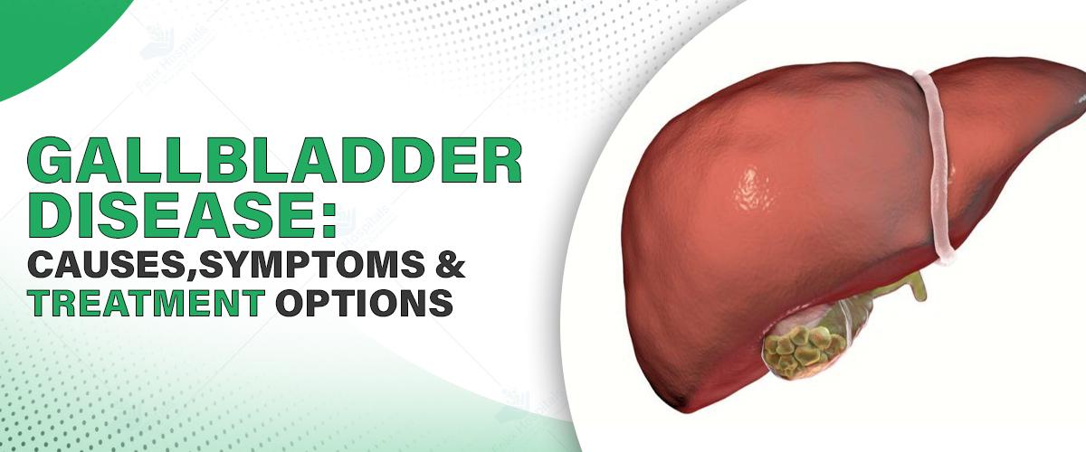
Gallbladder disease is a prevalent digestive disorder that usually arises from the existence of gallstones. Small, hardened bits of material that build up in the bile ducts can clog bile flow, leading to swelling, infection, and a lot of pain. The surgery to remove the gallbladder is usually the most extreme option. This blog shall offer a complete overview of gallbladder disease, its causes, signs, risk factors, treatment, and recovery. Before selecting a surgeon for gallbladder disease, consult the best laparoscopic treatment hospital in Noida for trusted advice.
Are you experiencing persistent abdominal pain or digestive discomfort? Don’t wait. Call us today at +91 9667064100.
The gallbladder is a small, pear-shaped organ lying under the liver. It has the main function of storing and releasing bile, a fluid produced by the liver that aids in the breakdown of fats in the small intestine. Gallbladder disease refers to several conditions that disrupt this organ's function, such as gallstones, inflammation, infections, and even cancer.
Gallbladder illness may be caused by the organ itself or by the bile ducts that are attached to it. As these ducts are common with other organs involved in digestion, any issue may be able to affect the liver, pancreas, and overall digestive health.
Gallstones (Cholelithiasis): The most common problem, gallstones are hardened bits created by extra cholesterol or bilirubin in bile. Some individuals can coexist with gallstones with no symptoms, while others can experience excruciating pain if a stone obstructs a bile duct.
Cholecystitis: Gallbladder inflammation, typically as a result of gallstones. Severe abdominal pain, fever, and vomiting result from acute cholecystitis. Scarring, gangrene, or rupture of the gallbladder may follow chronic cases.
Biliary Dyskinesia: The inability of the gallbladder to empty bile because of an issue with its motility. It is similar to gallstones and may need to be treated surgically.
Cancer of the Gallbladder: Although rare, gallbladder cancer can be a life-threatening diagnosis that is usually found later in its stages. Suspect growths should be surgically removed early.
Cholangiopathy: A collection of diseases of the bile duct, most often causing inflammation, infection, or constriction (strictures). Damage can impair both gallbladder and liver function for a long time.
Symptoms depend on the underlying cause but can consist of:
Severe pain in the upper right part of the abdomen (after meals)
Nausea and vomiting
Fever and chills
Jaundice (yellow color of the skin or eyes)
Dark-colored urine
Pale-colored stools
Digestive discomfort, bloating, or gas
These symptoms often appear suddenly and can last for several hours. It is important to get medical care in case of such signs.
Several risk factors are responsible for gallbladder disease:
High-cholesterol diet
Obesity (particularly with a BMI > 30)
Age ≥ 40 years
Female gender (due to hormonal factors such as estrogen)
Pregnancy or hormone replacement
Family history of gallbladder disease
Diabetes and Crohn's disease
Liver conditions such as cirrhosis
Certain ethnicities, particularly Native American or Mexican ethnicity
Physicians start with a careful symptom and medical history review. Physical examination can demonstrate abdominal pain, particularly in the upper right quadrant. Some of the diagnostic tests include:
Ultrasound: Initial imaging to identify gallstones and inflammation.
HIDA Scan: Assesses gallbladder function and bile flow.
Blood Tests: To identify infection, liver function, and inflammation.
CT Scan or MRI: For advanced imaging of the gallbladder and bile ducts.
Endoscopic Ultrasound (EUS) or ERCP: To identify and, at times, treat obstructions or remove gallstones.
Pain control and antibiotics are the initial interventions for inflammation or infection. These are often temporary measures, though.
In certain situations, an endoscope is employed to remove gallstones, insert stents, or drain abscesses, providing a minimally invasive alternative.
The most conclusive treatment is gallbladder removal, particularly in cases involving repeated gallstones or chronic cholecystitis. There are two main methods:
Laparoscopic Cholecystectomy: A minimally invasive procedure involving small cuts. Patients tend to recover rapidly and leave within 24–48 hours.
Open Surgery: Used for complicated conditions like severe infection, abscess, or gallbladder cancer.
Post-operative recovery differs with the surgery method:
Laparoscopic surgery usually enables a return to normal activity in 1 to 2 weeks.
Open surgery can take as much as 6 weeks to recover from.
Patients can have the following symptoms during the first few days after the surgery:
Abdominal pain or bloating
Changes in bowel habits, which are reversible
Fatigue
Dietary adjustments involve limiting fatty, greasy foods and eating high-fiber, easily tolerated foods. Most people adapt well and can resume a normal diet in a few months.
Untreated gallbladder disease can cause:
Severe infections (cholangitis, peritonitis)
Damage to the liver or cirrhosis
Pancreatitis (inflammation of the pancreas)
Rupture or gangrene of the gallbladder
Early diagnosis and treatment are critical to avoid long-term complications.
The cost of gallbladder removal surgery in India depends on:
Surgical technique (laparoscopic vs. open)
City and hospital facility
Hospital stay
Pre- and post-operative care
On average, gallbladder surgery in India will cost between ₹50,000 to ₹1.5 lakhs. Laparoscopic procedures will be a little higher but result in quicker recovery and fewer complications. It's better to approach the top laparoscopic surgeon in your city for a customized estimate.
You should consult the best laparoscopic surgeon in Noida if you experience any of the following conditions:
Recurring stomach pain, particularly after meals
Nausea or vomiting
Jaundice or fever
Loss of weight without reason
Early consultation leads to quicker relief and avoids complications.
Are you worried about the symptoms of your gallbladder or the cost of surgery? Schedule a consultation at the best hospital for gallbladder treatment.
Gallbladder disease may be prevalent, but it is incredibly curable if you catch it early. Thanks to today's diagnostic technologies and laparoscopic surgery, patients are back to their normal lives in no time. Always seek serious digestive issues and visit the top doctor or specialist in your area for professional examination and treatment. Take action on time, and your gallbladder—your life—may be saved.
Stay proactive. Listen to your gut. Your digestive health matters.
Q1. How long can someone live with gallstones without symptoms?
Ans: Many people live with silent gallstones for years without knowing it. If gallstones aren’t causing symptoms, they often don’t require treatment. However, regular monitoring is important because symptoms can develop unexpectedly, and complications like inflammation or infection can arise if left unchecked.
Q2. Is it possible to dissolve gallstones naturally or with diet changes?
Ans: In some cases, small cholesterol-based gallstones may be managed with medications like ursodeoxycholic acid, which can slowly dissolve them. However, this process is slow and doesn’t work for all stone types. Diet changes may reduce symptoms but rarely eliminate gallstones completely.
Q3. What dietary restrictions should be followed after gallbladder removal?
Ans: After gallbladder surgery, patients are usually advised to avoid fatty, fried, and greasy foods for a few weeks. Smaller, frequent meals help digestion. Over time, most people can return to a regular diet, though some may continue to experience mild food sensitivities, especially to high-fat items.
Q4. Can gallbladder disease cause issues with the pancreas or liver?
Ans: Yes, advanced gallbladder disease can affect the pancreas and liver. Gallstones may block the bile duct and cause bile to back up, leading to inflammation in the pancreas (pancreatitis) or liver damage. This makes early diagnosis and treatment critical to prevent further complications.
Q5. Are there any non-surgical treatments for gallbladder polyps?
Ans: Most small gallbladder polyps are benign and monitored with periodic ultrasounds. However, polyps larger than 1 cm or those that grow over time may pose a cancer risk and typically require surgical removal. Currently, there are no effective medications to shrink or treat polyps non-surgically.
Q6. What are the warning signs that gallbladder disease is becoming life-threatening?
Ans: Severe, persistent upper right abdominal pain, fever, yellowing of the skin or eyes (jaundice), vomiting, and confusion are red flags. These may indicate complications like gangrene, perforation, or widespread infection (sepsis). Immediate medical attention is required in such cases.

Understand the factors that influence gallbladder stone surgery cost in Noida, including hospital charges, surgeon expertise, type of procedure, and post-operative care expenses.

Expert gastroenterology care for better digestive health, offering advanced diagnosis and treatment for GERD, IBS, ulcers, and liver disorders.
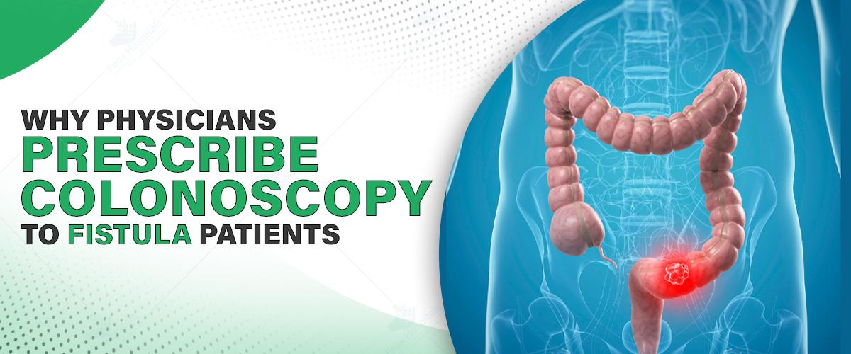
Anal and intestinal fistulas are abnormal tracts that form between two organs within the body or between an internal organ and the skin. These painful, persistent tracts can result in infection, drainage, and lifelong consequences if not diagnosed or treated. Although MRI and CT scanning are key in visualizing fistulas, colonoscopy continues to be essential in determining causes, establishing specific treatments, and enhancing patient outcomes.
Let’s explore why colonoscopy is frequently recommended for fistula patients and how it enhances the diagnostic and therapeutic process.
Ask your care team about scheduling a colonoscopy before imaging or surgery. Call us today at +91 9667064100.
The following ten factors support the doctor’s recommendation of colonoscopy for fistula surgery:
A good many fistulas are associated with underlying conditions such as Crohn's disease or ulcerative colitis, both inflammatory bowel diseases (IBD). External imaging may demonstrate the tract of a fistula, but colonoscopy enables physicians to directly view inflammation, ulcers, or mucosal abnormalities that establish a diagnosis of IBD.
Colonoscopy gives a complete view of large bowel, extending from the rectum to the cecum. This complete, real-time visualization helps in finding:
internal fistula openings
infections
structural abnormalities like scar tissue
polyps or precancerous signs
With the assistance of the best laparoscopic surgeon in Noida, polyps can be removed simultaneously, providing immediate preventative treatment against colorectal cancer.
What is found on colonoscopy has direct implications for the treatment of your fistula:
Straightforward fistulas: May be treated with local surgery like fistulotomy or seton insertion.
Complex or Crohn's fistulas: Typically require a multidisciplinary approach, utilizing a combination of biologic medications and surgery.
Suspicious findings, like polyps or lesions: Can involve further coordination with an oncologist or gastroenterologist.
This sensitivity ensures that each patient receives the right treatment at the right time, without inappropriate intervention.
Colonoscopy has also been demonstrated to enhance long-term results for fistula patients. A colonoscopy detects additional inflammation that is not visible on an external image. Treatment of these findings at the best laparoscopic surgery hospital in Noida can promote healing and lower the risk of recurrence of disease.
Fistulas can be associated with, or hide, severe conditions like colorectal cancer. Colonoscopy is the most effective method for early detection. Direct visualization and removal of precancerous lesions, like adenomatous polyps, are possible.
This type of examination is especially valuable in patients with chronic inflammation or rectal bleeding, where occult malignancy can otherwise be overlooked.
Colonoscopy findings frequently are the basis for a coordinated treatment plan among surgeons, gastroenterologists, and oncologists. In the case of complicated disease, this degree of coordination will ensure that every facet—ranging from controlling inflammation to operating timing—is managed in a smooth and patient-focused manner.
Done in mild sedation, colonoscopy is a safe, outpatient procedure with a rich harvest of diagnostic information. The process is more comfortable than anticipated in most patients, particularly when well-prepared.
The advantages of such a high-yield procedure—detection of disease, cancer prevention, and improved operative planning—far exceed the temporary inconvenience of bowel preparation.
Patients who are preparing for fistula repair can achieve bowel cleansing as part of their pre-operative routine. Colonoscopy aligns well with the process and allows providers to assess the bowel's readiness for repair. With instructions on the use of soothing creams, bidets, or soft wipes, discomfort during preparation can be reduced—making it more bearable even for patients with active fistulas.
Omitting colonoscopy prior to fistula surgery can lead to overlooked diagnoses or unsuccessful treatments. Surgeons might go ahead and operate without understanding the degree of internal inflammation or the presence of polyps or masses. Colonoscopy provides a detailed internal map—assisting in avoiding failed repairs or multiple operations.
For IBD or recurrent fistula patients, colonoscopy is not a standalone appointment—it's a part of ongoing monitoring of disease. Regular follow-ups allow evaluation of disease activity, assessment of drug efficacy, and identification of changes in the lining of the intestine that can predict future problems.
Schedule your colonoscopy today and take a proactive step toward full healing.
Colonoscopy is more than a cursory test for fistula patients—it's a thorough, diagnostic, and preventive procedure that enhances the outcome of surgery, individualizes care, and minimizes the risk of severe complications such as cancer or repeated surgery. When synchronized with the proper team of medical professionals, colonoscopy becomes a cornerstone of safe and effective fistula management.
From identifying hidden diseases to guiding complex treatment decisions, colonoscopy plays a central role in delivering the best possible care.
Q1. Is a colonoscopy necessary if an MRI already shows the fistula?
Ans: Yes. MRI maps the fistula tract, but colonoscopy can detect inflammation, ulcers, or polyps—critical details that an MRI cannot reveal.
Q2. Can colonoscopy worsen my fistula?
Ans: It's extremely uncommon. Done with caution under sedation, colonoscopy is safe. The majority of patients accept it well if they are well-prepared.
Q3. What happens if colonoscopy identifies Crohn's disease?
Ans: You could be placed on specific treatments such as biologic medications to manage inflammation prior to or following surgery—minimizing healing time and recurrence.
Q4. Will an oncologist be part of my treatment?
Ans: Only in the event that colonoscopy detects polyps or lesions that need follow-up evaluation. This facilitates early detection of cancer and planned intervention, if needed.
Q5. How much does a colonoscopy cost?
Ans: Although it does incur an added expense to overall care, early disease detection and prevention of repeated surgeries usually makes colonoscopy a cost-saving measure.
Q6. How often should I repeat colonoscopy if I have IBD?
Ans: For Crohn's or ulcerative colitis patients, repeat colonoscopies every 1–3 years as indicated, depending on disease severity and history.
Q7. Are there no-sedation colonoscopy options?
Ans: Yes, but the majority of patients like to have mild sedation for comfort. It relaxes the anxiety and enhances the quality and success of the procedure.

Anal fissures, perianal abscesses, and anal fistulas are common anorectal disorders that can significantly impact an individual's quality of life.

Cervical cancer is most common in women after breast cancer. Cervical cancer starts from the cervix, i.e., the cervix. The cervix is the lower part of the uterus that connects the vagina to the uterus.

Head and neck cancers are a heterogeneous group of cancers that can involve essential regions like the mouth, throat, voice box, and nasal passages. They are especially worrisome because of their proximity to vital organs and potential to affect simple activities such as speaking, eating, breathing, and making facial expressions. Head and neck cancers are treatable, however, even though serious, especially if found early.
This blog aims to provide a clear and comprehensive understanding of head and neck cancer, including its causes, symptoms, risk factors, and available treatment options. Raising awareness is crucial, as many early signs may seem minor or mimic common illnesses. However, consulting the best oncologist in Noida at the right time can lead to early diagnosis and timely treatment—factors that can make the difference between life and death.
If you have any recurring symptoms in your mouth, throat, or neck, get assessed promptly. Call us today at +91 9667064100.
Head and neck cancer involves various kinds of cancers that usually start in the squamous cells covering the moist surfaces of the head and neck. They are the mouth, throat (pharynx), nasal passages, voice box (larynx), and sinuses. A majority of them fall under the category of head and neck squamous cell carcinoma (HNSCC).
This group also includes less common cancers like those found in the salivary glands, which make saliva, and nasopharyngeal cancers, which are in the upper part of the throat behind the nose.
Oral Cancer – Occurs on the lips, gums, tongue, inner cheeks, roof of the mouth, and floor of the mouth.
Salivary Gland Cancer – Starts in the glands that secrete saliva.
Nasal Cavity and Sinus Cancer – Affected areas are in and around the nasal passages.
Nasopharyngeal Cancer – Develops in the upper throat.
Oropharyngeal Cancer – Develops in the middle throat, such as the tonsils and base of the tongue.
Hypopharyngeal Cancer – Develops in the lower throat.
Laryngeal Cancer – Specifically targets the voice box (larynx).
Symptoms depend on where the cancer is, but some warning signs are:
An ongoing sore throat
Pain or trouble swallowing
Hoarseness or a change in voice
Ear pain without infection
A neck or throat lump
A mouth or tongue sore that doesn't heal
Swelling in the jaw or face
Frequent nosebleeds or nasal blockage
Unintentional weight loss
Recurring headaches or facial pain
If you or your loved one is facing any symptoms like this so consult with the best oncologist in Noida for your healthier future.
Certain things make it more likely to get head and neck cancers:
Tobacco Use: The number one cause of head and neck cancer. Cigarettes, cigars, pipes, and smokeless tobacco.
Alcohol Use: Heavy drinking, particularly when combined with tobacco use, greatly raises risk.
Human Papillomavirus (HPV): Increasingly the cause of oropharyngeal cancers, particularly in younger adults.
Betel Nut Chewing: Prevalent in certain regions of Asia, highly associated with oral cancers.
Epstein-Barr Virus (EBV): Linked with nasopharyngeal cancer.
Radiation Exposure: History of previous radiation therapy to the head or neck.
Genetics: Genetic disorders such as Fanconi anemia.
Occupational Exposure: Wood dust, paint solvent, asbestos, and certain chemicals.
Poor Oral Hygiene: Associated with an increased risk of oral cancers.
Early detection is important. The procedure normally includes:
Physical Examination: Visual examination and palpation of the areas involved.
Endoscopy: Employment of a flexible tube with a camera to examine internal structures.
Imaging Tests: X-rays, CT scans, MRI, and PET scans to find tumors.
Biopsy: A tissue sample to determine the presence and type of cancer.
HPV Testing: Frequently completed when oropharyngeal cancer is suspected.
Staging tells the severity and how far it has spread. The TNM system is most often used:
T (Tumor): How big is the tumor, and where is
N (Node): How far the lymph nodes are involved
M (Metastasis): Whether it has spread to other parts of the body
Stages are I (early) through IV (advanced), determining treatment plans and outcomes.
Treatment is based on the type, location, and stage of cancer and can consist of a combination of:
1. Surgery
To remove the tumor and possibly surrounding lymph nodes. Minimally invasive methods are used when possible.
2. Radiation Therapy
External beam radiation targets the tumor directly. It can be used alone or in combination with surgery and chemotherapy.
3. Chemotherapy
Medications that destroy cancer cells or stop them from growing. Often combined with radiation therapy.
4. Targeted Therapy
Targets specific molecules that contribute to the growth of cancer. Cetuximab and Larotrectinib are two examples.
5. Immunotherapy
Activates the immune system to destroy cancer cells. Pembrolizumab and Nivolumab are two popular drugs.
6. Clinical Trials
New and experimental treatments are being tested.
7. Palliative Care
Manages symptoms and enhances quality of life during treatment, particularly in late stages.
Treatments for head and neck cancer can impact speech, swallowing, and appearance. Supportive care can include:
Reconstructive surgery
Nutritional support
Psychological counseling
You can reduce your risk of head and neck cancers by:
Avoiding tobacco and secondhand smoke
Restricting alcohol consumption
Receiving the HPV vaccine
Practicing good oral hygiene
Using sun protection
Avoiding contact with harmful chemicals
Make an appointment for a consultation if you observe anything out of the ordinary. Knowledge is the beginning of protection.
Head and neck cancers are a broad spectrum of disorders, which, although serious, may be successfully treated, particularly when caught early. A chronic sore throat, swallowing trouble, or unusual lumps are symptoms that should never be dismissed. Early detection combined with an appropriate treatment regimen can result in much better outcomes and quality of life.
Prevention through healthy lifestyle practices—such as quitting smoking, avoiding alcohol, and taking the HPV vaccine—can significantly lower your risk. If you've already been diagnosed, it's most important to know about your treatment options. If you're in quest of getting insights into the cost of head and neck cancer treatment in Noida, there is a need to find out those options that provide state-of-the-art care, multidisciplinary treatment, and transparent pricing to enable you to plan accordingly.
Knowing better, doing better, and paying attention to early warning signs can be a lifesaver. Don't wait—take control of your health and seek the care you need.
Q1. Can head and neck cancer result in signs that resemble a cold?
Ans: Yes, initial signs are often subtle, such as a sore throat or hoarseness, and are wrongly assumed to be less severe illnesses.
Q2. Can head and neck cancer be developed without tobacco and alcohol consumption?
Ans: Yes. HPV-related head and neck cancer is becoming more prevalent among non-smokers and non-users of alcohol.
Q3. Can dental problems be an early indicator of oral cancer?
Ans: Yes, the presence of ongoing mouth sores, bleeding gums, or poorly fitting dentures can be symptoms.
Q4. Does HPV-related throat cancer manifest differently?
Ans: HPV-related head and neck cancers tend to occur with a neck lump and little pain, unlike classical symptoms.
Q5. Is the risk of head and neck cancer present in young adults?
Ans: Yes, particularly because of increasing HPV infections. The cancers are no longer limited to older adults.
Q6. Can head and neck cancer recur after treatment?
Ans: Yes, recurrence can happen, particularly in the first two years. Routine follow-up is important.
Q7. Do these cancers affect mental health?
Ans: Yes. The observable and functional consequences of treatment can affect self-esteem and emotional well-being.

There has been a steep rise in hearing issues in the last 5 years. People who use earphones and headphones continuously for long duration are prone to become deaf.

Chronic neck pain can limit daily life—consult a spine expert today for accurate diagnosis and effective treatment options.

Explore essential information about breast cancer, including its symptoms, causes, types, and treatments, to stay informed and proactive about your health.

Recent studies have explored a possible link between frequent mouthwash use and cancer risk.
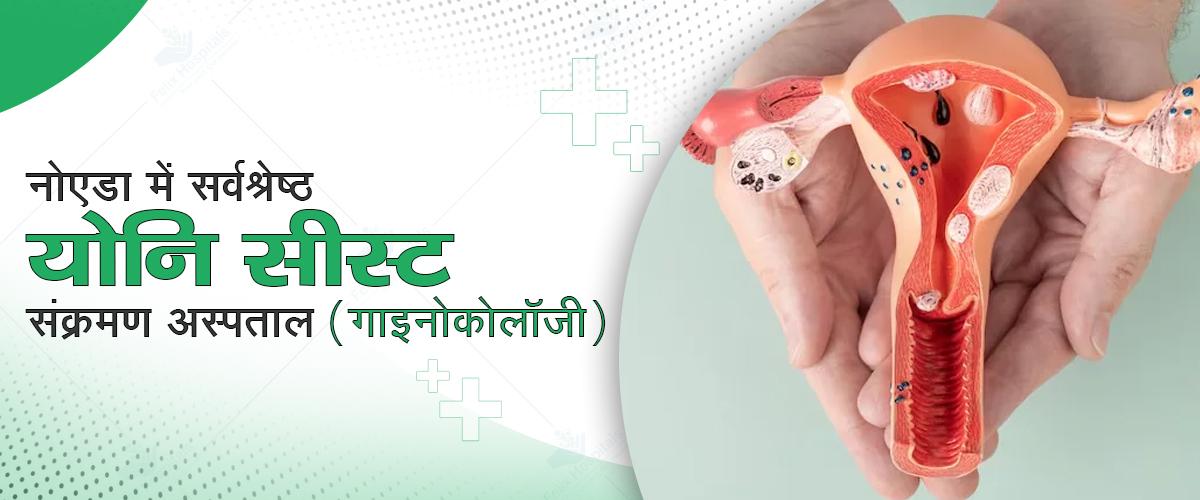
भागदौड़ भरी जिंदगी में महिलाएं सेहत को लेकर लापरवाह रहती हैं। खासतौर पर जब बात निजी अंगों की हो, तो महिलाओं में योनि संक्रमण (Vaginal Infection) और योनि सीस्ट (Vaginal Cyst) जैसी समस्याएं बेहद आम हैं। मगर शर्म और जागरूकता की कमी के कारण इनका सही समय पर इलाज नहीं हो पाता, जो बाद में गंभीर स्वास्थ्य समस्याओं का कारण बन सकता है। इस ब्लॉग में हम योनि संक्रमण, विशेष रूप से यीस्ट इंफेक्शन के बारे में जानकारी साझा करेंगे — इसके लक्षण, कारण, और बचाव के उपाय।
अगर आप इन समस्याओं का सही और सुरक्षित इलाज ढूंढ रही हैं, तो नोएडा में सर्वश्रेष्ठ गाइनोकोलॉजी हॉस्पिटल का चुनाव करना बेहद जरूरी है, जहां विशेषज्ञ डॉक्टरों की देखरेख में आधुनिक जांच और इलाज की सुविधा उपलब्ध हो।
योनि सीस्ट एक छोटी, मुलायम व तरल-से भरी गांठ होती है, जो महिला की योनि की आंतरिक या बाहरी दीवार में बनती है। यह सीस्ट बिना लक्षण के होती हैं। महिला को पता भी नहीं चलता कि वह इसके साथ जी रही है। जब सीस्ट में संक्रमण होता है तब यह दर्द, सूजन और दुर्गंधयुक्त डिस्चार्ज जैसी समस्याएं पैदा करती है। सरल भाषा में कहे तो योनि सीस्ट एक तरह की गांठ है, जो संक्रमित होने के बाद इन्फेक्टेड सीस्ट बनती है। योनि सीस्ट में संक्रमण तब होता है जब बैक्टीरिया या अन्य सूक्ष्म जीव उसमें घुसते हैं। इसके कारणों योनि की स्वच्छता में लापरवाही, असुरक्षित यौन संबंध, जन्म या प्रसव के दौरान चोट, बार-बार का इनफेक्शन या एंटीबायोटिक का अत्यधिक उपयोग होता है।
सबसे आम प्रकार योनि के दोनों ओर नीचे की तरफ बार्थोलिन ग्रंथियां होती हैं, जो लुब्रिकेशन में मदद करती हैं। जब इस ग्रंथि का छिद्र बंद होता है, तो तरल अंदर इकट्ठा होने से सीस्ट बनती है।
भ्रूण के विकास के दौरान बनने वाली संरचनाओं से यह सीस्ट बनती है, यह योनि की साइड वॉल में बनती है। यह अधिकतर बिना लक्षण होती है, लेकिन बड़ी होने पर असुविधा होती है।
यह अक्सर प्रसव या शल्य चिकित्सा के दौरान चोट से बनती है। योनि की भीतरी परत के नीचे की स्किन जब बढ़ती है, तो गांठ होती है। यह छोटी होती हैं किन दर्द का कारण बनती हैं।
अनदेखी एक सामान्य सीस्ट को गंभीर संक्रमण में बदलती है। योनि सीस्ट में संक्रमण होने के पीछे कई कारण होते हैं। यहां निम्न चार कारण प्रमुख हैं:
लैक्टोबैसिलस हानिकारक जीवाणुओं को नियंत्रित रखते हैं। जब यह संतुलन बिगड़ता है, तो कैनडीडा अल्बिकन्स जैसी फंगस तेजी से बढ़ती है, इससे फंगल इंफेक्शन होता है। गार्डनेरेला वेजिनेलिस जैसे बैक्टीरिया के बढ़ने से बैक्टीरियल वेजिनोसिस होता है।
बिना कंडोम के यौन संबंध बनाना या बार-बार पार्टनर बदलना योनि संक्रमण की वजह है। इससे न केवल एसटीआई यानी गोनोरिया, क्लेमाइडिया, ट्राइकोमोनास का खतरा बढ़ता है, साथ ही संक्रमण फैलकर सीस्ट में भी पहुंचता है।
महिलाओं के शरीर में हार्मोन खासकर एस्ट्रोजन और प्रोजेस्टेरोन प्रजनन क्रिया को नियंत्रित करते हैं। यदि किसी कारण जैसे तनाव, पीसीओडी, थायरॉइड रोग या मेनोपॉज हार्मोन असंतुलित होते हैं तो ग्रंथियों की कार्यप्रणाली बिगड़ती है, इससे ग्रंथियों का तरल बाहर नहीं निकल पाता और सीस्ट बनती है।
गलत तरीके से साफ-सफाई, अधिक रगड़ या गंदे अंडरगारमेंट्स पहनने से समस्या होती हैं। योनि क्षेत्र में बैक्टीरिया या फंगस का जमाव पसीने और नमी की वजह से संक्रमण होता है। इस कारण सीस्ट की त्वचा पर सूक्ष्म चोट से बैक्टीरिया अंदर जा सकते हैं।
योनि सीस्ट में जब संक्रमण होता है, तो कुछ लक्षण स्पष्ट रूप से उभरते हैं। यदि इन संकेतों को समय पर पहचान लिया जाए, तो गंभीर समस्या बनने से पहले इलाज संभव होता है।
योनि के आसपास हल्की सूजन, एक छोटी सी गांठ होती है। शुरुआत में यह दर्दरहित होती है, लेकिन संक्रमण बढ़ते ही यह गर्म, लाल और स्पर्श में दर्दनाक होती है। इस कारण चलने, बैठने या यौन संबंध बनाते समय असहजता महसूस होती है।
योनि क्षेत्र में लगातार जलन या चुभन जैसा महसूस होती है, खासकर जब सीस्ट में मवाद भरता है। बैठने, टाइट कपड़े पहनने या चलते समय यह दर्द बढ़ता है।
योनि से निकलने वाला डिसचार्ज यदि पीला, हरा या बदबूदार हो, तो यह संक्रमण का सीधा है। संक्रमण की स्थिति में यह डिसचार्ज गाढ़ा और चिपचिपा होता है।
जब सीस्ट का आकार या सूजन मूत्रमार्ग के पास पहुंचती है, पेशाब करते समय जलन या तेज दर्द होता है, बार-बार पेशाब इसके लक्षण है, कभी-कभी महिलाएं इसे यूटीआई समझती हैं, लेकिन यह अंदरूनी सीस्ट संक्रमण का प्रभाव होता है।
हर महिला में योनि संक्रमण या सीस्ट का खतरा होता है, लेकिन कुछ विशेष वर्ग की महिलाओं में यह जोखिम होता है।
बार-बार यौन संबंध बनाना या अनसेफ सेक्स करना संक्रमण की संभावना बढ़ाता है। एसटीआई (यौन संचारित संक्रमण) के संपर्क में आने से योनि की ग्रंथियां संक्रमित होती हैं।
मधुमेह के कारण शरीर की रोग प्रतिरोधक क्षमता कमजोर होती है। छोटी-सी सीस्ट में भी संक्रमण तेजी से फैलता है, घाव भरने में अधिक समय लगता है।
बार-बार दवाइयों का सेवन योनि के नेचुरल बैक्टीरियल बैलेंस को बिगाड़ता है। इससे संक्रमण और फंगल ग्रोथ को मौका मिलता है जिससे सीस्ट अधिक संवेदनशील होती है।
गर्भावस्था के दौरान हार्मोनल बदलाव, पसीना, नमी और इम्यूनिटी में बदलाव से योनि क्षेत्र संक्रमण के प्रति अधिक संवेदनशील होता है। सीस्ट गर्भावस्था को प्रत्यक्ष प्रभावित नहीं करती, लेकिन दर्द से जीवन गुणवत्ता जरूर गिरती है।
अगर लक्षण महसूस हो तो अगला जरूरी कदम है सटीक जांच। डॉक्टर आमतौर पर निम्नलिखित जांच कराते हैं:
डॉक्टर हाथ से या स्पेकुलम की मदद से योनि क्षेत्र की जांच करते हैं। गांठ की स्थिति, आकार, रंग और दर्द की जानकारी की जाती है।
योनि या पेट के जरिए अल्ट्रासाउंड कर सीस्ट की गहराई, आकार और स्थिति का पता लगाते हैं। इससे पता चलता है कि सीस्ट तरल-भरी है या ठोस है।
अगर डिस्चार्ज होता है, तो उसे लैब में जांचते हैं जिससे यह पता चले कि उसमें कौन-सा बैक्टीरिया या फंगस है। इससे यह आसान होता है कि कौन-सी दवा संक्रमण के लिए प्रभावी होगी।
अगर सीस्ट असामान्य दिखे या लंबे समय तक ठीक न हो तो उसकी एक छोटी सी परत लेकर लैब टेस्ट करते हैं। कैंसर जैसे गंभीर खतरे की संभावना को जांचने के लिए होता है।
समय पर इलाज न केवल दर्द से राहत देता है, बल्कि संक्रमण के फैलने से रोकता है। योनि सीस्ट संक्रमण के इलाज के प्रमुख तरीके निम्न है:
यदि सीस्ट में बैक्टीरियल संक्रमण है, तो डॉक्टर टॉपिकल एंटीबायोटिक्स देते हैं। मगर दवा कभी भी डॉक्टर की सलाह के बिना न लें। वहीं, वार्थिन डक्ट मार्सुपियलाइजेशन विशेष रूप से बार्थोलिन सीस्ट के लिए इस्तेमाल की जाने वाली एक प्रभावी प्रक्रिया है। इसमें डॉक्टर सीस्ट को काटकर उसका तरल बाहर निकालते हैं, फिर उसमें कैथेटर लगाते हैं ताकि वह दोबारा न बने और संक्रमण दोहराया न जाए।
इस प्रक्रिया के प्रकार, अस्पताल की सुविधाओं और डॉक्टर की विशेषज्ञता पर निर्भर करती है। आमतौर पर यह लागत कुछ हजार से लेकर कुछ दस हजार रुपये तक हो सकती है। सटीक जानकारी के लिए किसी विश्वसनीय गाइनोकोलॉजी सेंटर या अस्पताल से संपर्क करना उचित होगा।
योनि सीस्ट संक्रमण में थोड़ी सतर्कता और अच्छी आदतों से रोका जा सकता है।
रोजाना गुप्तांग की सफाई करें, खुशबूदार साबुन, डियोड्रेंट या वेजाइनल वॉश से परहेज करना चाहिए, यह पीएच बैलेंस बिगाड़ते हैं, मासिक धर्म के दौरान हर चार–छह घंटे में पैड/टैम्पोन बदलें, नहाने के बाद योनि को सुखाएं, जिससे नमी से संक्रमण नहीं हो।
सूती अंडरगारमेंट्स पसीना सोखते हैं। टाइट, नायलॉन या सिंथेटिक अंडरवियर से नमी बनती है, जो फंगल और बैक्टीरियल संक्रमण को न्योता देती है इसलिए रोजाना अंडरगारमेंट्स बदलना चाहिए।
कंडोम का इस्तेमाल संक्रमणों से सुरक्षा देता है। बार-बार पार्टनर बदलना या बिना जांच के संबंध बनाना एसटीआई यौन संचारित संक्रमण का खतरा बढ़ाता है। असुरक्षित यौन संबंध सेहत के लिए खतरा है।
हर 6–12 महीने में एक बारजांच कराना चाहिए। जिससे पहले से मौजूद सीस्ट, फाइब्रॉइड या इन्फेक्शन का समय पर पता चल सके।
योनि संक्रमण की अनदेखी करना गंभीर परिणाम देता है। एक छोटी सी गांठ या हल्का दर्द भी किसी बढ़ते संक्रमण का संकेत होता है। जो आगे चलकर शारीरिक कष्ट का कारण बनता है। प्रजनन क्षमता और मानसिक स्वास्थ्य पर असर डालता है। महत्वपूर्ण यह नहीं कि संक्रमण हुआ या नहीं, बल्कि यह है कि आपने कब ध्यान दिया जाए। नोएडा जैसे विकसित शहर में अनुभवी गाइनोकोलॉजिस्ट, आधुनिक तकनीकें और गोपनीय इलाज के विकल्प उपलब्ध हैं।
योनि सीस्ट संक्रमण को लेकर अक्सर पूछे जाने वाले सवाल और उनके जवाब (Frequently asked questions and answers about vaginal cyst infection)
प्रश्न 1: क्या योनि सीस्ट खतरनाक होती है ?
उत्तर: अधिकांश योनि सीस्ट बिना किसी लक्षण के होती हैं, जो अपने-आप ठीक होती हैं। अगर उनमें दर्द, सूजन या मवाद हो तो इलाज जरूरी है।
प्रश्न 2 : योनि सीस्ट संक्रमण से बांझपन होता है ?
उत्तर: आमतौर पर सीस्ट बांझपन का कारण नहीं होती है। मगर संक्रमण फैल जाए और समय पर इलाज न हो, तो यह गर्भाशय या फैलोपियन ट्यूब को प्रभावित करता है। जिससे प्रजनन क्षमता प्रभावित होती है।
प्रश्न 3: क्या योनि सीस्ट खुद से फूट सकती है ?
उत्तर: कुछ मामलों में संक्रमित सीस्ट फूटती है। मवाद बाहर निकलता है। यह दर्दनाक होता है। संक्रमण आसपास के ऊतकों में फैलता है।
प्रश्न 4: क्या मासिक धर्म (पीरियड्स) पर सीस्ट का असर पड़ता है ?
उत्तर: सीधे तौर पर नहीं लेकिन अगर सीस्ट बड़ी हो या उसमें सूजन होती है। यह पीरियड्स के दौरान दर्द और असहजता बढ़ती है।
प्रश्न 5: बार्थोलिन सीस्ट और अन्य सीस्ट में क्या अंतर है ?
उत्तर: बार्थोलिन सीस्ट योनि के प्रवेश द्वार के पास होती है। बार्थोलिन ग्रंथि के बंद होने से बनती है। अन्य सीस्ट जैसे गार्टनर डक्ट या इन्क्लूजन सीस्ट अलग कारणों से बनते हैं।
प्रश्न 6: क्या योनि सीस्ट कैंसर में बदलती है ?
उत्तर: दुर्लभ मामलों में योनि सीस्ट कैंसर का रूप लेती है। लेकिन अगर सीस्ट बार-बार हो तो डॉक्टर द्वारा बायोप्सी कराना जरूरी है।
प्रश्न 7: क्या गर्भावस्था के दौरान सीस्ट का इलाज संभव है ?
उत्तर: दवाएं और इलाज केवल वहीं अपनाए जाते हैं जो गर्भवती महिला और भ्रूण दोनों के लिए सुरक्षित हों। सर्जरी केवल जरूरत पड़ने पर ही की जाती है।

पीसीओडी (PCOD) की समस्या उन महिलाओं में अधिक देखने को मिल रही है, जो नाइट शिफ्ट में काम करती हैं। पूरी रात जागना देर रात खाना जैसी लाइफस्टाइल उनकी सेहत को भारी नुकसान पहुंचा रही है।

Sexually transmitted infection is one of the significant public health problems. And the bigger problem is people hide sexual dysfunction instead of discussing it.

डिमेंशिया व्यक्ति की स्मृति, सोचने-समझने की क्षमता, निर्णय लेने और व्यवहार पर गहरा असर डालता है। यह रोग धीरे-धीरे प्रगति करता है और समय के साथ व्यक्ति की दैनिक गतिविधियों को पूरी तरह से प्रभावित कर सकता है। डिमेंशिया (Dementia) को समय रहते समझना और इसके विभिन्न चरणों की पहचान करना इलाज और देखभाल की दृष्टि से अत्यंत आवश्यक है।
इस ब्लॉग में हम डिमेंशिया के कारण, प्रमुख लक्षण, और विभिन्न चरणों पर विस्तार से चर्चा करेंगे, ताकि आप या आपके किसी प्रियजन को शुरुआती संकेतों को पहचानने में मदद मिल सके।
अगर आप डिमेंशिया की जांच (Dementia testing), उपचार या देखभाल को लेकर किसी विशेषज्ञ से सलाह लेना चाहते हैं, तो नोएडा में सर्वश्रेष्ठ हॉस्पिटल से संपर्क करें, जहां अनुभवी न्यूरोलॉजिस्ट (Neurologist) और आधुनिक सुविधाएं उपलब्ध हैं। अधिक जानकारी के लिए हमसे संपर्क करें — समय पर उठाया गया एक कदम, जीवन की दिशा बदल सकता है।
अपॉइंटमेंट शेड्यूल करने के लिए, अभी कॉल करें: +919667064100
डिमेंशिया में व्यक्ति की याददाश्त, सोचने-समझने की शक्ति, भाषा, निर्णय क्षमता और व्यवहार धीरे-धीरे क्षीण होती हैं। यह कोई एकल बीमारी नहीं है होती बल्कि लक्षणों का एक समूह है, जो मस्तिष्क की सामान्य कार्यप्रणाली में रुकावट उत्पन्न करता है। डिमेंशिया की सबसे सामान्य वजह अल्जाइमर रोग है। 65 वर्ष से अधिक आयु के लगभग 5-8% लोग किसी न किसी प्रकार के डिमेंशिया से प्रभावित होते हैं डिमेंशिया को एक प्रगतिशील न्यूरोडीजेनेरेटिव डिसऑर्डर (Neurodegenerative Disorders) के रूप में देखा जाता है। जिसमें समय के साथ मस्तिष्क कोशिकाएं नष्ट होती जाती हैं। समय रहते इसका निदान और देखभाल रोग की गति को धीमा कर सकती है। रोगी के जीवन की गुणवत्ता को बेहतर बना सकती है।
अल्जाइमर रोग (Alzheimer's disease) डिमेंशिया का सबसे सामान्य कारण है। इसमें मस्तिष्क में प्रोटीन जमा होने से कोशिकाएं क्षतिग्रस्त हो जाती हैं। वेस्कुलर डिमेंशिया में मस्तिष्क में रक्त प्रवाह बाधित होने से होता है। इससे सोचने की क्षमता और ध्यान केंद्रित करने में दिक्कत होती है। लूई बॉडी डिमेंशिया में मस्तिष्क की नसों में असामान्य प्रोटीन जमा होते हैं। इसमें भ्रम, नींद की दिक्कतें और संतुलन की समस्याएं होती हैं। फ्रंटोटेम्पोरल डिमेंशिया के कारण मस्तिष्क के सामने और किनारे के हिस्से में क्षति होती है। व्यक्ति के व्यवहार, भाषा और निर्णय लेने की क्षमता पर असर पड़ता है। लंबे समय तक पार्किंसन होने के बाद कई लोगों को डिमेंशिया हो सकता है। सिर की चोट, मस्तिष्क में संक्रमण, शराब या नशीले पदार्थों का लंबे समय तक सेवन, विटामिन बी 12 की कमी, थायरॉइड या लिवर से जुड़ी समस्याएं, डिप्रेशन भी इसके कारणों में शामिल हैं।
अल्जाइमर रोग (Alzheimer's disease):
इसमें मस्तिष्क में एमायलॉइड प्लाक्स और टॉ प्रोटीन का असामान्य जमाव होता है। जिससे स्मृति, भाषा और सोचने की क्षमता प्रभावित होती है। धीरे-धीरे बढ़ने वाला यह रोग प्रारंभ में व्यक्ति को हाल की बातें याद रखने में भूलाता है।
वैस्कुलर डिमेंशिया (Vascular Dementia)
यह तब होता है जब मस्तिष्क में रक्त प्रवाह बाधित होता है। इसमें निर्णय लेने की क्षमता, एकाग्रता और योजना बनाने में कठिनाई होती है, लक्षण अचानक भी उभरते हैं, हर स्ट्रोक के बाद स्थिति और बिगड़ सकती है।
लुई बॉडी डिमेंशिया (Lewy Body Dementia)
इसमें मस्तिष्क की तंत्रिका कोशिकाओं में लुई बॉडी नामक असामान्य प्रोटीन जमा होते हैं। इसके लक्षण अल्जाइमर और पार्किंसन (Parkinson's) दोनों से मिलते-जुलते हैं, इसमें भ्रम की स्थिति, नींद से संबंधित विकार, चलने में कठिनाई और दृष्टि से जुड़ा भ्रम होता है।
फ्रंटोटेम्पोरल डिमेंशिया (Frontotemporal dementia)
यह डिमेंशिया मस्तिष्क के फ्रंटल और टेम्पोरल लोब्स को प्रभावित करता है। जो व्यक्ति के व्यवहार, निर्णय क्षमता और भाषा को नियंत्रित करते हैं। यह 40–65 वर्ष की आयु के बीच शुरू होता है। इसमें रोगी में व्यवहार में बदलाव, सामाजिक अनुशासन की कमी और बोलने में समस्या होती है।
चरण 1 – सामान्य कार्यशीलताः
कोई भी स्मृति हानि या मानसिक समस्या नहीं होती है। इसमें मस्तिष्क सामान्य रूप से काम करता है, व्यवहार और संज्ञानात्मक क्षमता सामान्य होती है।
चरण 2 – बहुत हल्की हानिः
कभी-कभी सामान्य भूलने की घटनाएं होती है जैसे चाबी या चश्मा रखकर भूल जाना। उम्र से जुड़ी सामान्य बातें यानी कोई स्पष्ट रोग संकेत नहीं होता है।
चरण 3 – हल्का संज्ञानात्मक अवक्रमणः
ध्यान केंद्रित करने में कठिनाई होती है, निर्णय लेने में संकोच होता है, सामाजिक कार्यों में परेशानी महसूस होती है, इस चरण में परिवार वालों को फर्क महसूस होता है
चरण 4 – मध्यम संज्ञानात्मक हानिः
स्पष्ट लक्षण सामने आते हैं जैसे तिथि, स्थान, पैसे का हिसाब याद न रखना। जटिल कार्यों यानी बिल भरना, बैंकिंग में असमर्थता होती है।
चरण 5 – मध्यम गंभीर हानिः
दैनिक जीवन की गतिविधियों में मदद की जरूरत पड़ती है जैसे कपड़े पहनना, खाना बनाना, नहाना आदि। अपने घर का पता या परिवार के सदस्यों के नाम भूलना।
चरण 6 – गंभीर संज्ञानात्मक गिरावटः
व्यक्ति को अपने परिजनों के नाम याद नहीं रहते हैं। व्यवहार में बदलाव, भ्रम, रोना या गुस्सा आना, रात में बेचैनी, नींद में गड़बड़ी, टॉयलेट जैसी गतिविधियों में भी सहारे की आवश्यकता होती है।
चरण 7 – बहुत गंभीर स्थितिः
बोलने में कठिनाई, शब्दों की कमी होती है, चलने-फिरने में असमर्थता होती है, निगलने में दिक्कत, पूर्ण देखभाल की आवश्यकता होती है। यह चरण पूर्ण शारीरिक और मानसिक निर्भरता का होता है
डिमेंशिया का इलाज तभी प्रभावी हो सकता है जब उसका समय पर निदान हो। इसके लिए न्यूरोलॉजिस्ट कई स्तरों पर जांच करते हैं:
डिमेंशिया या अन्य न्यूरोलॉजिकल समस्याओं की पहचान के लिए रोगी की संज्ञानात्मक क्षमता, स्मृति, संतुलन, बोलने, और सोचने की क्षमता की विस्तृत जांच की जाती है। इस प्रक्रिया में डॉक्टर यह तय करते हैं कि मस्तिष्क के कौन-से न्यूरोलॉजिकल फंक्शन प्रभावित हो रहे हैं और उनकी गंभीरता क्या है।
इस तरह की गहन जांच और सही निदान के लिए आपको अनुभवी विशेषज्ञ की आवश्यकता होती है। यदि आप किसी भरोसेमंद विशेषज्ञ की तलाश में हैं, तो नोएडा में अच्छे न्यूरोलॉजिस्ट से संपर्क करना एक सही कदम हो सकता है। यहां कई reputed हॉस्पिटल्स में अनुभवी न्यूरोलॉजिस्ट उपलब्ध हैं, जो आधुनिक तकनीकों और जांच विधियों की मदद से सटीक इलाज सुनिश्चित करते हैं।
मस्तिष्क की संरचना में बदलाव जैसे मस्तिष्क संकोचन, स्ट्रोक या ट्यूमर का पता लगाते हैं। यह डिमेंशिया के अन्य संभावित कारणों को बाहर करने में मदद करता है।
इसमें स्मृति, ध्यान, गणना, भाषा और कॉन्शसनेस की जांच की जाती है। स्कोर के आधार पर डिमेंशिया की गंभीरता का निर्धारण करते हैं।
थायरॉइड, विटामिन बी 12 की कमी आदि ब्लड जांच यह सुनिश्चित करती है कि संज्ञानात्मक गिरावट किसी अन्य इलाज योग्य समस्या के कारण तो नहीं हो रही है।
डिमेंशिया से पूरी तरह बचाव की कोई गारंटी नहीं है, लेकिन जीवनशैली में बदलाव और कुछ सावधानियों से इसके जोखिम को काफी हद तक कम किया जा सकता है।
रोजाना कम से कम 30 मिनट पैदल चलें, योग या हल्का व्यायाम करें। व्यायाम मस्तिष्क में रक्त संचार को बेहतर बनाता है।
माइंड डाइट और मेडिटेरेनियन डाइट अपनाएं। हरी सब्जियां, फल, नट्स, मछली, जैतून का तेल, साबुत अनाज का सेवन करें, तले-भुने, प्रोसेस्ड फूड और ज्यादा नमक-शक्कर से परहेज करें।
दिमागी खेल खेलें यानी सुडोकू, पहेली, ताश, शतरंज, नई चीजें सीखें यानी नई भाषा, संगीत वाद्य, लेखन या पेंटिंग करें।
रक्तचाप, कोलेस्ट्रॉल और डायबिटीज को नियंत्रण में रखें क्योंकि जो दिल के लिए अच्छा है, वही दिमाग के लिए भी अच्छा है।
धूम्रपान और शराब से दूरी बनाएं ये मस्तिष्क कोशिकाओं को नुकसान पहुंचाते हैं और डिमेंशिया का खतरा बढ़ाते हैं।
नींद पूरी और अच्छी लें, हर रात 7–8 घंटे की गहरी नींद लें, नींद में रुकावट या स्लीप एप्निया जैसी समस्याओं का इलाज करवाएं।
तनाव को नियंत्रित करें योग, ध्यान, प्राणायाम और सामाजिक मेलजोल तनाव कम करने में सहायक हैं।
सुनने की क्षमता का ध्यान रखें, सुनाई न देना डिमेंशिया के खतरे को बढ़ाता है जरूरत हो तो हियरिंग एड का इस्तेमाल करें।
सामाजिक रूप से जुड़े रहें अकेलेपन से मानसिक रोगों का खतरा बढ़ता है। परिवार, दोस्तों के साथ समय बिताएं।
नियमित जांच कराएं विशेषकर यदि पारिवारिक इतिहास हो तो साल में एक बार न्यूरोलॉजिस्ट से परामर्श लें (Consult a neurologist)।
डिमेंशिया का उपचार केवल दवाओं तक सीमित नहीं होता। इसका व्यापक प्रबंधन परिवार, देखभाल, वातावरण और जीवनशैली पर भी निर्भर करता है।
औषधीय प्रबंधनः
डोनेपेजिल (अल्जाइमर रोग के शुरुआती से मध्यम चरणों में प्रयोग में लाई जाती है। मेमंटीन मध्यम से गंभीर डिमेंशिया में सहायक होती है, रिवास्टिगमाइन मस्तिष्क में रसायन संतुलन बनाए रखने में मदद करती है, गैलेंटामाइन और अन्य अल्जाइमर-स्पेसिफिक दवाएं इलाज में सहायक होती है। यह दवाएं लक्षणों में सुधार ला सकती हैं, लेकिन रोग को पूरी तरह से रोक नहीं सकतीं।
थैरेपी आधारित हस्तक्षेपः
संज्ञानात्मक व्यवहार थेरेपी (CBT) भ्रम, चिंता और अवसाद जैसे मानसिक लक्षणों को संभालने में काफी सहायक होती है। यह थेरेपी मरीज को नकारात्मक सोच को समझने और सकारात्मक सोच विकसित करने में मदद करती है।
मेमोरी ट्रेनिंग में स्मृति को बेहतर करने के लिए विशेष गतिविधियां और अभ्यास कराए जाते हैं, जो डिमेंशिया या अल्जाइमर से जूझ रहे मरीजों के लिए लाभदायक होते हैं।
वहीं, म्यूजिक और आर्ट थेरेपी रचनात्मक अभिव्यक्ति के माध्यम से मानसिक संतुलन बनाए रखने में मदद करती है। यह थेरेपी न केवल तनाव को कम करती है, बल्कि रोगी को भावनात्मक रूप से जोड़े रखने और सामाजिक रूप से सक्रिय बनाने में भी उपयोगी होती है।
जहां तक बात है नोएडा में इलाज की कीमत की, तो यह विभिन्न थेरेपी सेशन्स की संख्या, हॉस्पिटल या क्लिनिक की सुविधा, और विशेषज्ञ की फीस पर निर्भर करती है। आम तौर पर CBT और मेमोरी ट्रेनिंग की प्रति सेशन कीमत ₹1,000 से ₹3,000 तक हो सकती है, जबकि आर्ट और म्यूजिक थेरेपी के पैकेज अलग-अलग हो सकते हैं। सटीक जानकारी के लिए किसी विश्वसनीय क्लिनिक या अस्पताल से संपर्क करना उचित होगा।
परिवार और देखभाल प्रबंधनः
डिमेंशिया रोगी के लिए दवाओं की समय पर निगरानी, पोषण, मानसिक सहयोग और दैनिक जरूरतों की पूर्ति में सक्रिय भागीदारी जरूरी होती है। घर का माहौल सुरक्षित और सरल बनाना भी जरूरी होती है। इससे स्पष्ट संकेतक, पर्याप्त रोशनी और न्यूनतम विघटन यानी रूटीन बनाना रोगी को मानसिक रूप से स्थिर बनाता है। रोगी को अपनापन, धैर्य और सम्मान देना बेहद जरूरी है। उनकी बातों को महत्व देना, उन्हें निर्णय में शामिल करना आत्मविश्वास बढ़ाता है।
रोग की प्रगति और जीवनशैलीः
हल्का व्यायाम जैसे टहलना, योग मानसिक स्थिति में सुधार लाता है। संतुलित आहार विशेषकर मेडिटेरेनियन डाइट यानी फल, सब्जी, फाइबर, ओमेगा-3 मस्तिष्क को पोषण देता है। बोर्ड गेम्स, पहेली, ड्राइंग, किताबें पढ़ना मस्तिष्क को सक्रिय रखते हैं। संगीत सुनना और पुराने फोटो देखना सकारात्मक स्मृतियों को सक्रिय करता है। पूरा आराम और अच्छी नींद संज्ञानात्मक स्वास्थ्य के लिए अनिवार्य है। रिश्तेदारों, दोस्तों या ग्रुप गतिविधियों से जुड़े रहना मानसिक गिरावट को कम करता है।
डिमेंशिया में व्यक्ति की याददाश्त, सोचने की क्षमता, निर्णय लेने की क्षमता और दैनिक कार्यों को करने की क्षमता प्रभावित होती है। लक्षण नजर आने पर जल्द से जल्द डॉक्टर से मिलना जरूरी होता है। न्यूरोलॉजिस्ट (Neurologist) डॉक्टर मस्तिष्क और तंत्रिका तंत्र के रोगों के विशेषज्ञ होते हैं। वह डिमेंशिया की सटीक जांच, ब्रेन स्कैन, न्यूरोलॉजिकल परीक्षण और दवा प्रबंधन में सबसे अहम भूमिका निभाते हैं।
अभी अपॉइंटमेंट शेड्यूल करें – कॉल करें: +91 9667064100
डिमेंशिया एक जटिल लेकिन पहचाने जाने वाला न्यूरोलॉजिकल विकार है। जिसे समय रहते पहचानना और देखभाल देना बेहद जरूरी है। यह केवल स्मृति की समस्या नहीं, बल्कि जीवन की गुणवत्ता को गहराई से प्रभावित करने वाला रोग है। अगर न्यूरोलॉजी गाइडलाइन के अनुसार समय पर निदान और उपचार शुरू किया जाए, तो इसकी प्रगति को धीमा किया जा सकता है। चुनौती से निपटने के लिए परिवार, समाज और चिकित्सा प्रणाली तीनों की मिलीजुली भागीदारी जरूरी है। समझ, सहानुभूति और वैज्ञानिक उपायों से हम डिमेंशिया से प्रभावित लोगों को सक्रिय जीवन जीने में सहायता कर सकते हैं।
प्रश्न 1. डिमेंशिया के कितने चरण होते है ?
जवाब: डिमेंशिया को सात चरणों में बांटा गया है। जिन्हें वैश्विक गिरावट पैमाना (जीडीएस) के अनुसार वर्गीकृत करते हैं। यह चरण हल्की भूलचूक से लेकर गंभीर मानसिक और शारीरिक निर्भरता तक बढ़ते हैं।
प्रश्न 2. क्या डिमेंशिया का इलाज संभव है ?
जवाब: डिमेंशिया का पूर्ण इलाज संभव नहीं है। मगर दवाओं, थैरेपी और जीवनशैली प्रबंधन से इसके लक्षणों को नियंत्रित कर सकते हैं। रोग की गति को धीमा कर सकते हैं।
प्रश्न 3. डिमेंशिया का पता कैसे चलता है ?
जवाब: डिमेंशिया का निदान न्यूरोलॉजिस्ट संज्ञानात्मक परीक्षण, न्यूरोलॉजिकल एग्जामिनेशन, एमआरआई/सीटी स्कैन और ब्लड टेस्ट शामिल होते हैं।
प्रश्न 4. डिमेंशिया किन लोगों को अधिक होता है ?
जवाब: यह रोग सामान्यत 65 वर्ष से अधिक उम्र के लोगों में पाया जाता है। कुछ मामलों में यह 40-60 की उम्र में भी शुरू होता है।
प्रश्न 5. डिमेंशिया और भूलने की बीमारी में क्या अंतर है ?
जवाब: सामान्य भूलने की आदतें उम्र का हिस्सा हो सकती हैं, लेकिन डिमेंशिया में यह भूलने की प्रवृत्ति रोजमर्रा के जीवन को बाधित करती है। इसमें लोगों को पहचानना, रास्ता भूल जाना या बातों को बार-बार दोहराना शामिल होता।
प्रश्न 6. क्या डिमेंशिया के रोगी ठीक हो सकते हैं ?
जवाब: समय पर निदान हो और समुचित देखभाल मिले तो रोगी कई वर्षों तक संतुलित और सम्मानजनक जीवन जी सकता है।
प्रश्न 7. क्या डिमेंशिया के रोगियों को अकेला छोड़ना ठीक है ?
जवाब: डिमेंशिया के रोगी को हमेशा देखरेख की जरूरत होती है। अकेलेपन से उनकी स्थिति और बिगड़ती है। इसलिए परिवार या देखभालकर्ता की भूमिका अहम होती है।
प्रश्न 8. क्या डिमेंशिया में भ्रम और गुस्सा सामान्य हैं ?
जवाब: जैसे-जैसे रोग बढ़ता है। मरीजों में भ्रम, चिड़चिड़ापन, व्यवहार परिवर्तन और रात में बेचैनी होती हैं। इन्हें थैरेपी, वातावरण और संयम से नियंत्रित कर सकते हैं।

ब्रेन स्ट्रोक एक जानलेवा स्थिति है जिसमें मस्तिष्क को रक्त आपूर्ति रुक जाती है। इसके प्रकार, कारण, लक्षण, बचाव और इलाज

पार्किंसंस रोग एक न्यूरोलॉजिकल विकार है, जो मस्तिष्क के कुछ हिस्सों में डोपामाइन नामक रसायन के स्तर में कमी के कारण होता है।

सीजर (Seizure) जिसे हिंदी में दौरे कहा जाता है, एक ऐसी स्थिति है जिसमें मस्तिष्क की कोशिकाओं (न्यूरॉन्स) की असामान्य विद्युत गतिविधि के कारण शरीर पर अचानक और अनियंत्रित झटके, अजीब हरकतें या बेहोशी जैसे लक्षण उत्पन्न होते हैं।

माइग्रेन (Migraine) के लक्षण, कारण और निवारण के उपाय जानें। सही समय पर पहचान और देखभाल से सिरदर्द और असुविधा कम होती है

Experiencing repeated episodes of loose motion? It's worse than just discomfort—it can ruin your health if not treated in time. Many residents of Noida search for "the best hospital near me" to get instant and effective relief from such gastrointestinal disorders. Loose motion, or what is commonly called diarrhea, could be an indication of numerous underlying conditions, ranging from food intolerance to infection.
Understanding when to go to the doctor and whom to go to can be the difference between life and death. In this blog, we are going to be discussing the causes, diagnosis, and treatment of loose motion, and acquainting you with the top general physicians in Noida who specialize in treating this disorder.
Call us today at +91 9667064100 to schedule your appointment and begin the path to better digestive health!
Loose motion is defined by frequent watery stools. Although an occasional diarrhea is harmless, frequent or chronic loose motion can cause dehydration, weakness, and even hospitalization if left untreated. Based on the duration and severity, loose motion can be:
Acute: It persists for a short duration of three days and is most often due to infection or food poisoning.
Chronic: It persists for more than two weeks and can be indicative of an underlying serious disease.
Loose motion is due to numerous diseases, such as:
Gastroenteritis: Due to bacterial or viral infection.
Irritable Bowel Syndrome (IBS): Functional gastrointestinal disorder.
Lactose Intolerance: Milk product undigestion inability.
Inflammatory Bowel Disease (IBD): Crohn's disease and ulcerative colitis fall in this category.
Food Poisoning: Due to rotten food or water.
Parasitic Infections: Mostly occur in unhygienic environments.
If you get loose motions regularly, it is better to see an expert to eliminate severe gastrointestinal illness.
Identification of the reason behind loose motion is important to deal with it properly. As per the top internal medicine department of Noida, some of the most common reasons are:
Infections: Viral (e.g., rotavirus), bacterial (e.g., E. coli, Salmonella), or parasitic infections.
Contaminated Food or Water: Particularly during travel or monsoon.
Food Allergies or Intolerances: For instance, gluten or lactose intolerance.
Medications: Antibiotics may upset gut bacteria and produce diarrhea.
Digestive Disorders: For instance, IBS, IBD, or celiac disease.
Stress and Anxiety: May upset digestion and cause loose motions.
To find out the underlying cause, physicians typically do:
Physical Examination: Evaluation of hydration status and tenderness in the abdomen.
Stool Tests: Identification of infection or parasites.
Blood Tests: Identification of inflammation or electrolyte imbalance.
Endoscopy or Colonoscopy: In chronic and/or red-flag presenting patients, e.g., in blood in stools.
Food Allergy Tests: Where there is intolerance.
Early diagnosis makes treatment possible in terms of prevention of unnecessary complications.
Treatment is either in a pattern or recognition of pattern:
Oral Rehydration Therapy (ORT): Should restore the lost electrolytes and fluid.
Antibiotics or Antiparasitics: For treating established bacterial or parasitic infection.
Probiotics: Help restore normal gut flora.
Antidiarrheal Medicines: Like loperamide, for temporary relief.
Dietary Adjustments: Bland diet, avoiding dairy and high fiber foods initially.
Treatment of Underlying Disease: Such as treating IBS or IBD if diagnosed.
Physicians recommend avoidance of self-medication, especially if there is fever, blood in stool, or long-standing diarrhea.
In case of digestive issues, the best general physician in Noida or an internal medicine specialist is usually your first point of contact. They examine your symptoms, conduct necessary tests, and refer you to a gastroenterologist if required. For residents in and around Noida, consulting a reliable doctor at a well-known clinic ensures timely and accurate treatment.
Felix Hospital in Noida is famous for its modern-day treatment and well-experienced internal medicine doctors. In case you experience frequent loose motions, the following physicians provide professional diagnosis and empathetic treatment:
Dr. Anshumala Sinha – Famous for her accurate diagnosis and effective medical treatment of gastroenterological conditions.
Dr. Neelabh Pratap – Provides well-sustained treatment regimens and patient-focussed treatment of stomach conditions.
Dr. Priyanka Singh – Experienced in treating acute and chronic loose motion causes.
Dr. Ravi Sharma – Experienced in internal medicine with expertise in gastrointestinal well-being.
Dr. Sonakshi Saxena – Experienced in treating infection and food causes of diarrhea.
Dr. Keshav Kumar Garg – Experienced in evidence-based treatment of gastrointestinal and metabolic disorders.
Dr. Apoorva Shetty – Specialist in treating complicated gastrointestinal symptoms and patient-friendly solutions.
These doctors provide not only medical expertise but also guidance on preventive healthcare and lifestyle modification to prevent recurring attacks.
Visit our experienced doctors at Felix Hospital in Noida for the correct diagnosis and customized treatment schedule. Call us at +(91) 9667064100 for appointments and more information.
Loose motion might look like a mere irritation, but if it becomes repetitive or catches up with you some time down the line, it might be an omen indicating more sinister gastric issues. Early specialist consultation is important in diagnosis of cause and to avoid complications like dehydration or malnutrition. Good and affordable care, with awareness of loose motion cost in Noida and opting for a reputed hospital like Felix, can create peace of mind and normal healing.
The best way of managing loose motion is through believing in veteran doctors who do not only treat but also provide long-term counsel. With skilled doctors at Felix Hospital, you can take command of your lifestyle and digestive system.
Q1. What is causing loose motion on a regular basis?
Ans: Recurrent loose motion may have various causes such as infection (bacterial, viral, or parasitic), intolerance to food, gastrointestinal disorder like IBS, food poisoning, or stress and anxiety. The specific cause will be decided by consulting a doctor.
Q2. How is loose motion diagnosed?
Ans: Physicians diagnose loose motion through physical examinations, stool and blood tests, and sometimes sophisticated examinations such as endoscopy or colonoscopy, particularly when symptoms recur or are severe. Diagnosis determines treatment.
Q3. When do I need to see a doctor for loose motion?
Ans: If you have loose motions frequently, dehydration, hematemesis, or symptoms for more than a few days, it is mandatory that you see a doctor. Long or excessive diarrhea can cause severe health problems.
Q4. What is the best medicine for loose motion?
Ans: Loose motion treatment varies with its cause. In bacterial or parasitic infections, antibiotics or antiparasitic drugs can be given. Oral rehydration, probiotics, and dietary modifications are usually advised. In long-standing diseases such as IBS or IBD, a prolonged treatment regimen may be required.
Q5. Is loose motion treatment costly in Noida?
Ans: The price of treating loose motion in Noida depends on the reason behind the condition and diagnostic tests needed. But clinics such as Felix provide quality but affordable treatment for chronic as well as acute loose motion, providing high-class treatment at reasonable rates.
Q6. Does loose motion lead to dehydration?
Ans: Yes, repeated loose motion might cause dehydration that, if left untreated, can be perilous. Oral fluids or IV fluids to rehydrate and replace lost fluids and electrolytes are usually required.
Q7. Who is the best doctor for treatment of loose motion in Noida?
Ans: Some of the best general physicians and best gastroenterologists in Noida are available at Felix Hospital, including Dr. Anshumala Sinha, Dr. Neelabh Pratap, Dr. Priyanka Singh, and Dr. Ravi Sharma. They are known for offering effective loose motion treatment in Noida, providing precise diagnoses and personalized treatment plans for quick recovery.

Gastric problems occur when excess acid or digestive disturbances affect the stomach lining, causing symptoms like bloating, acidity, and pain. Timely diagnosis ensures effective treatment.
Expert gastroenterology care for better digestive health, offering advanced diagnosis and treatment for GERD, IBS, ulcers, and liver disorders.
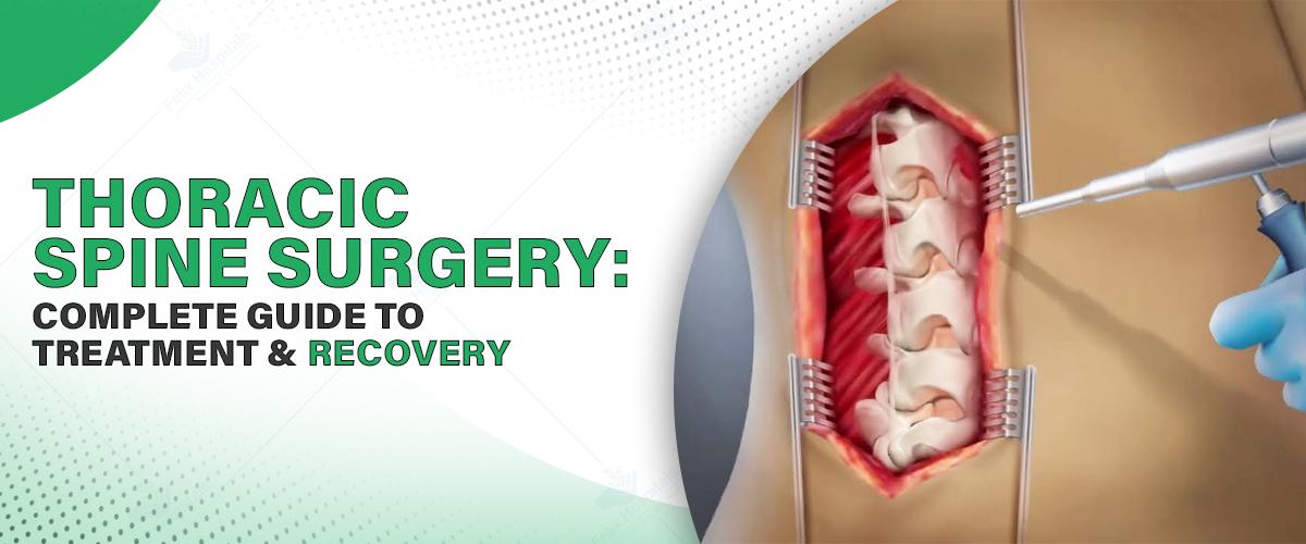
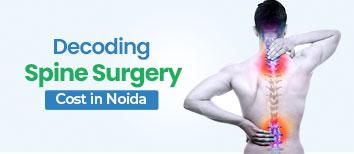
The thoracic spine is the central part of the human spinal column, located between the cervical (neck) and lumbar (lower back) areas. These parts of the spine are responsible for stability, mobility, and guarding organs. For maintenance of the spine and timely medical intervention, it is crucial to know about its anatomy, function, associated conditions, diagnosis, and treatment. Finding the best thoracic spine surgery in Noida is crucial, so contact the nearest hospital with the best reputation in your area and set up your consultation right away.
Book your consultation by calling +91 9667064100 with a spine specialist today and take the first step toward relief.
There are 12 vertebrae in the thoracic spine, labeled T1-T12. It starts at the end of the neck and goes up to just below the lumbar spine, constituting the upper and middle back. They protect the spinal cord and support the rib cage. The thoracic spine is less flexible than the cervical or lumbar spine, thus less prone to injury; however, it can still develop disorders.
Each thoracic vertebra is connected to a rib pair, making up a rigid thoracic cage that protects the heart and lungs. There is an intervertebral disc located between each vertebra, acting as a shock absorber, offering flexibility. Facet joints in this area control and limit spinal movements, enabling proper functioning.
The thoracic spine has some important roles:
Protection: It surrounds the spinal cord and shields nerves that report to organs like the lungs, heart, liver, and intestines.
Support: It stabilizes the rib cage and upper portion to support posture and movement.
Respiration: The configuration of the thoracic area and muscles that are connected to it makes breathing possible by opening up and closing the chest.
Mobility and Stability: While the thoracic spine has little flexion and extension, it also has the greatest rotational motion of any of the spinal segments with stability.
Postural Alignment: It is also responsible for maintaining the normal curve of the spine, which assists with balance and the transmission of forces effectively.
Twelve pairs of spinal nerves exit the thoracic spine:
T1-T2: The nerves impact the upper chest and arms. The T1 nerve is one of the nerves that belong to the brachial plexus, responsible for controlling hand and arm movement.
T3-T5: Regulate chest wall, lungs, and diaphragm.
T6-T12: Contribute support to abdominal and back muscles and help in balance, posture, and actions such as coughing and breathing.
These nerves transmit impulses to and from large organs such as the lungs, heart, liver, kidneys, and small intestine.
While injuries to the thoracic spine are less common given its stiffness, there are still various conditions that will impact it:
Muscle Strain or Irritation: Most often caused by poor posture or prolonged sitting.
Ligament Sprain: Due to sudden twisting motions or trauma.
Spinal Tumors: Benign or malignant tumors that often present as deep, chronic pain at night.
Vertebral Compression Fractures: Most common in elderly or osteoporotic individuals, with resultant pain and loss of height.
Overuse Injuries: Due to repetitive use like lifting, twisting, or bending.
Spinal Fractures: Typically secondary to trauma or bone weakening by osteoporosis or tumors.
With age or wear and tear, the thoracic spine can develop the following:
Spondylosis (Osteoarthritis): Cartilage wear and tear leads to pain and stiffness.
Degenerative Disc Disease: Discs that separate the vertebrae dehydrate and become less elastic.
Spinal Stenosis: Narrowing of the spinal canal that may crush the spinal cord and nerves.
Kyphosis: Excessive forward spinal curve that causes slouching posture.
Herniated Discs: It might occur in the thoracic spine and may press on spinal nerves.
Myelopathy: Compression of the spinal cord that impacts mobility and coordination.
Thoracic nerve damage may lead to:
Radiating pain or numbness closer to the chest or belly
Weakness of limbs or core muscles
Shortness of breath or difficulty breathing
Loss of bowel or bladder control
Decreased sensation in arms, legs, or genitalia
Stiffness or spasms of the muscle
If you do experience these symptoms, ensure you see the best spine surgeon in Noida as soon as possible.
The accurate diagnosis begins with the history and physical examination. The ideal physician can recommend:
X-rays: To observe bone alignment and breaks.
MRI: To examine the spinal cord, discs, and nerve roots.
CT Scan: Gives detailed pictures of vertebrae and structural issues.
Myelogram: Injects contrast dye into the spinal cord and nerves, highlighting them.
EMG and Nerve Conduction Studies: Test for nerve function and identify locations of compression.
Treatment differs by severity and cause. The most common options are:
Physical Therapy: Trims and adjusts posture.
Medications: Muscle relaxants, pain medications, and anti-inflammatories.
Epidural Steroid Injections (ESIs): End nerve pain and decrease inflammation.
Bracing: Helps stabilize the spine when it has fractures or kyphosis.
Decompression Surgery: Removes bone or tissue pressing on nerves.
Spinal Fusion: Merges the spine by rods, screws, and bone grafts.
Tumor Removal: Requires delicate surgical procedures to eliminate tumors from the spine.
The best thoracic spine hospital in Noida has an expert who will prescribe the optimal course depending on the health and the complexity of the condition.
Recovery could include rehabilitation therapies such as
Physical Therapy: Restores strength and mobility.
Occupational Therapy: Helps to adapt to daily routines.
Pain Management: Medication and Non-Invasive Therapies.
Lifestyle Modifications: Posture improvement, weight management, and ergonomic adjustments.
Follow-ups and imaging studies on a regular basis assist in monitoring progress. Follow-up is usually based on the severity of the condition, timely intervention, and patient compliance.
Seek immediate care if you have:
Increasing or persistent back pain
Numbness or weakness in the legs or arms
Loss of bowel or bowel control
Chest pain or shortness of breath
Unexplained weight loss accompanied by back pain
These could be signs of life-threatening conditions like compression, fracture, or tumor in the spinal cord that should be examined immediately at a hospital.
The cost of treating thoracic spine disorders is determined by the following factors:
Diagnostic tests (MRI, CT scan, X-rays)
Treatment process (physical therapy, surgery, medication)
Hospitalization (number of days) and room
Recovery and follow-up after treatment
In India, the cost of treatment ranges from ₹50,000 to ₹5 lakhs or more, depending on how complicated the condition is, the technology used, and the amount of experience of the specialist. Always ask for an estimate from the hospital.
The thoracic spine surgery is instrumental in stabilizing the human body, shielding important organs, and facilitating movement. Pathologies in this area can lead to pain, loss of mobility, or systemic issues if not treated. An early diagnosis, a consultation with a competent spine specialist, and prompt treatment at a well-reputed hospital can significantly enhance the quality of life.
If you’re experiencing persistent upper back discomfort, posture issues, or unusual neurological symptoms, consult a spine specialist today. Prioritize spinal health—get evaluated at the best hospital nearby for expert care and peace of mind.
Q1. Can poor posture alone cause permanent thoracic spine problems?
Ans: Yes, repeated poor posture—especially slouching or forward head position—over time will cause structural changes of the thoracic spine, including kyphosis, muscle imbalance, and premature degenerative disc disease.
Q2. Why does thoracic spine pain worsen at night or with recumbency?
Ans: Thoracic pain increases at night due to decreased distraction of the spinal joints and changes in intervertebral disc pressures. It may also reflect the development of tumors or infections of the spine that require urgent examination.
Q3. Can thoracic spine problems cause heart- or lung-type symptoms?
Ans: Yes. Distressed thoracic nerves can mimic tightness or pain in the chest, commonly mistaken for heart or lung problems. This symptom is very common with thoracic radiculopathy or dysfunction of the costovertebral joint.
Q4. Is exercise with thoracic disc bulging or herniation safe?
Ans: Exercise is generally alright, but twisting or high-impact activity must be avoided without seeking expert guidance. A spine specialist or physiotherapist can come up with a safe regime.
Q5. How do I differentiate thoracic nerve pain from muscular back pain?
Ans: Thoracic nerve pain will radiate around the rib cage in a band-like manner and is sometimes accompanied by generalized tingling or numbness. The pain will generally be localized to the affected area and worsen with specific movements or pressure.
Q6. Can hours of sitting at a desk cause thoracic spine degeneration?
Ans: Yes. Sedentary living and poor ergonomics over time contribute largely to degenerative thoracic changes, especially if compounded by inadequate back and core muscle strength.

Spinal cord injury is one of the most serious injuries one can have. The damages can be permanent if not treated on time.

Advanced neurosurgery improves treatment for brain and spine disorders with better outcomes.

Spinal stenosis surgery is needed when severe pain, weakness, or numbness persists despite non-surgical treatment and affects daily activities.
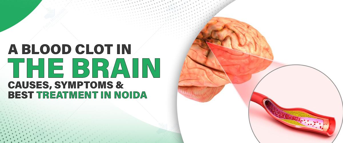
We all get headaches. Sometimes from stress, dehydration, or staring at a screen too long. But if a headache feels more intense and more sudden, especially followed by confusion, blurry vision, or difficulty speaking, it needs immediate medical attention. It might be a sign of something much more serious like a blood clot in brain.
Therefore, understanding what causes a blood clot, how to spot it, and most importantly, what doctors can do about it, can make all the difference. Timely treatment saves lives – and in many cases, prevents long-term damage.
So if you or a loved one experiences a sudden bout of headaches, confusion, or weakness in your face or limbs, don’t brush it off. These could be early signs of something serious like a brain clot. Seek medical help immediately!
At Felix Hospital, our neurosurgeons are trained to respond quickly with advanced diagnostics and treatment. To schedule an appointment, contact us at +(91) 9667064100!
A blood clot in the brain, also known as a cerebral thrombosis or stroke caused by thrombosis, happens when blood flow to part of the brain gets blocked by a clot. The brain, like every other organ, needs oxygen and nutrients. When a clot stops that flow, brain cells can start to die within minutes.
There are different types of strokes caused by clots. The most common is an ischemic stroke, which is due to a blockage. This is different from a hemorrhagic stroke, which involves bleeding in the brain.
Sometimes, the clot forms in a brain artery itself. Other times, it travels from another part of the body, like the heart or neck, and gets stuck in a narrow artery in the brain.
Clots don’t usually form out of nowhere. They often result from other underlying issues. Here are some of the most common causes of brain blood clot:
High blood pressure – Damages blood vessel walls, making clots more likely
Diabetes – Affects how your blood flows and how well your vessels work
Atherosclerosis – Hardening or narrowing of arteries due to plaque buildup
Atrial fibrillation – An irregular heartbeat that can cause clots in the heart to travel to the brain
Smoking – Increases clotting tendency
High cholesterol – Leads to blocked arteries over time
Obesity and sedentary lifestyle – Raise overall risk of stroke and clot formation
Head trauma – In some cases, injury to blood vessels can lead to clot formation
When a blood clot blocks an artery in your brain, symptoms usually come on suddenly. You might feel fine one moment, and very unwell the next. Common signs of blood clot in brain include:
• A sudden, severe headache
• Numbness or weakness, especially on one side of the body
• Difficulty speaking/slurred speech
• Trouble understanding what others are saying
• Blurred or double vision
• Dizziness or loss of balance
• Confusion or trouble concentrating
• Loss of consciousness or seizures
• F – Face drooping
• A – Arm weakness
• S – Speech difficulty
• T – Time to call emergency services
Doctors rely on a combination of symptoms and advanced imaging to figure out what’s going on. The process usually involves:
CT Scan or MRI: These help to identify if there’s a clot or bleeding.
CT Angiography or MR Angiography: These focus on the blood vessels and show exactly where the blockage might be.
Blood Tests: To check clotting factors, sugar levels, and cholesterol
ECG and Echocardiogram: If there’s a chance the clot came from the heart, these tests help detect heart conditions
If you suspect signs of a blood clot in brain, acting fast is the key! The treatment depends on how quickly the person gets to the hospital. The golden window is often the first 3 to 4.5 hours.
Thrombolytic (Clot-Busting) Drugs : The medication works by dissolving the clot and restoring blood flow. It must be given within a few hours of symptom onset to be effective.
Mechanical Thrombectomy: In some cases, especially with larger clots, doctors use a catheter to go into the brain’s arteries and physically remove the clot. This is done under imaging guidance and is highly effective when performed early.
Blood Thinners: If the stroke risk remains high, patients may be put on long-term anticoagulants to prevent future clots.
Lifestyle Changes and Rehabilitation: Post-stroke treatment isn’t just about medication. Physical therapy, occupational therapy, and speech therapy play a big role in recovery, especially if mobility or communication was affected.
A blood clot in the brain is serious and should be treated as a medical emergency. In many cases, it’s preventable. As mentioned, rapid treatment can dramatically change the outcome. At Felix Hospital, we provide comprehensive treatment for brain blood clots, including emergency care. We also provide rehabilitation and support care to all our patients recovering from the after effects of the brain blood clot or brain stroke.
Don’t ignore early signs and symptoms, especially if your body is giving you warning signs. Consult the best neurologists in Noida immediately to seek appropriate treatment.
Q1. Can you survive a brain clot?
Ans: Yes, many people survive and recover well, especially with early treatment. The key is recognizing the signs and getting help fast.
Q2. How long is the recovery after a brain clot?
Ans: It varies. Some recover in weeks, while others may need several months of rehab. Age, overall health, and how quickly treatment was received all play a role.
Q3. Are blood clots in the brain preventable?
Ans: Often, yes. Managing blood pressure, quitting smoking, staying active, and keeping diabetes under control can significantly lower your risk.
Q4. What’s the difference between a stroke and a brain clot?
Ans: A stroke caused by a clot is called an ischemic stroke. So technically, a brain clot is one type of stroke. There are other kinds too, like hemorrhagic strokes, which involve bleeding.
ब्रेन स्ट्रोक एक जानलेवा स्थिति है जिसमें मस्तिष्क को रक्त आपूर्ति रुक जाती है। इसके प्रकार, कारण, लक्षण, बचाव और इलाज

A concise guide explaining brain tumor surgery cost in Noida, including key factors, treatment options, and expert care.

Recognizing the symptoms of a brain stroke is critical for getting prompt medical attention. The acronym F.A.S.T. is often used to remember the most common Brain stroke symptoms.

Brain-boosting foods rich in antioxidants, omega-3s, and vitamins support memory, protect brain cells, and may help reduce the risk of dementia and Alzheimer’s disease.
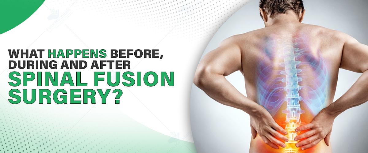
Spinal fusion surgery is a standard process applied to correct different spine disorders like herniated disks, spinal instability, scoliosis, or degenerative disc disease. Through spinal fusion surgery, the goal is to attach two or more vertebrae in your back permanently, removing any motion between them to avoid pain and enhance stability. If you are looking for the best spinal fusion physicians near me, learning about this surgery in detail will help you make informed choices. This blog will prepare you for spinal fusion surgery by explaining what to expect.
Meet the best orthopedic surgeon at the start. Call us now at +91 9667064100.
1. First Consultation and Assessment: The process starts with an initial consultation with a spine expert. The spine expert will assess your symptoms, medical history, and imaging studies such as X-rays, MRIs, or CT scans to decide whether spinal fusion is appropriate for your case. The spinal fusion operation price in Noida varies based on the level of complexity and hospital amenities.
2. Preoperative Instructions: The following suggestions may be included in the preoperative instructions:
Quit smoking, as it affects bone healing.
Avoid taking anti-inflammatory medications that can lead to increased bleeding.
Make arrangements for assistance at home while you are recovering.
Get the required blood work and physical examination.
3. Pre-Surgery Preparation One day before the surgery
You may be requested to:
Fast for a minimum period of 8 hours before surgery
Take a shower using antibacterial soap
Pack a hospital bag with essentials for your hospital stay, such as your medical records and comfortable clothes
1. Anesthesia and Positioning: The surgery is done under general anesthesia. Depending on the part of your spine being operated on, you will lie on your stomach or back.
2. The Surgical Process
The surgeon makes incisions over the affected area.
The surgeon removes any ruptured disc material.
A bone graft (either from your body or a donor) is inserted in between vertebrae.
Metal rods, screws, or plates might be attached to stabilize the spine.
Spinal fusion surgery can last several hours, depending on how many vertebrae are fused.
1. Hospital Stay and Recovery Room After surgery: You will be taken to the recovery room, where your vital signs are monitored closely. Most patients are hospitalized for 2 to 4 days. In this process, doctors monitor pain management and the resumption of mobility safely.
2. Mobility and Pain Management
Pain medications are administered to reduce discomfort.
Physical therapists can get you moving within 24 hours.
Gradual walking and basic movements are encouraged to prevent stiffness.
3. Healing and Long-Term Recovery
The fusion process typically takes 6 to 12 months.
You should avoid bending, twisting, or lifting heavy objects.
We may provide a back brace for additional support.
Follow-up visits are necessary to monitor healing and make adjustments in care.
As with any major surgery, spinal fusion comes with certain risks, including:
Infection
Nerve damage
Incomplete fusion (pseudoarthrosis)
Pain at the bone graft site
Talking to your specialist about these risks will allow you to balance the advantages against possible complications.
Decreases chronic back pain
Stabilizes the spine
Enhances mobility and posture
Allows patients to go back to regular activities with improved comfort
Most patients can return to work and physical activities within a few months with proper rehabilitation and medical attention.
Finding the correct team for your treatment is critical. If you are in search of a spinal fusion hospital in Noida, keep the following in mind:
Surgeon experience
Presence of high-end surgical equipment
Facility reputation
Transparency of charges and patient feedback
Selecting treatment at the best Orthopedic hospital in Greater Noida provides you with multidisciplinary teams, post-operative services, and custom treatment plans.
Schedule an appointment with the best doctor in Noida for personalized care and post-operative care.
Spinal fusion is an established way to treat long-term spinal disorders and enhance the quality of life. Be it assessing your options or getting ready for the surgery, having the proper information at hand can assist you in going through the process with ease. If you are looking to undergo the procedure, begin by taking advice from experts at the best hospital in Greater Noida.
From diagnosis to recovery, skilled care ensures you receive the treatment, protection, and support you require for your healing journey.
Q1. What is the recovery time from spinal fusion surgery?
Ans: Recovery can take between 3 to 12 months, depending on patient health, age, and activity level. Physical therapy and routine follow-up are important aspects of recovery.
Q2. Is spinal fusion surgery painful?
Ans: Post-surgery pain is normal, but it is well-managed with medication and improves over time.
Q3. Can spinal fusion fail?
Ans: Infrequently, but sometimes, pseudoarthrosis or non-union occurs, where the two bones do not join together properly. This can be minimized with great surgical skill and good postoperative care.
Q4. Will I become less mobile in my back after spinal fusion?
Ans: Spinal fusion restricts motion between two fused vertebrae but generally does not limit overall mobility. Most patients adjust easily.
Q5. What is the average cost of spinal fusion surgery in Noida?
Ans: It varies with complexity, hospital, and fees of the surgeon. It usually ranges between ₹1.5 lakhs and ₹4 lakhs. Always seek a proper estimate before going for surgery.
Spinal stenosis surgery is needed when severe pain, weakness, or numbness persists despite non-surgical treatment and affects daily activities.
A spinal cord injury occurs when there is damage to the spinal cord, often resulting from trauma, disease, or degenerative conditions.
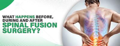
Spinal fusion surgery is a procedure that joins two or more vertebrae in the spine to reduce pain, correct deformity, or improve stability.

Subscribe to our
© Copyright 2026. All Rights Reserved by Felix Healthcare Private Limited
Design and development by :


 +(91)9667064100
 Emergency : +(91)9667064100