MRI Scan
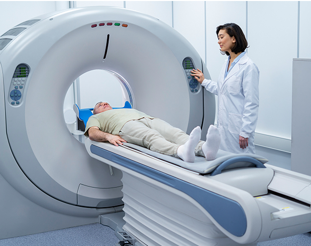
Magnetic resonance imaging (MRI) is a medical imaging procedure that produces precise images of the organs and tissues in your body through the magnetic field and computer-generated radio waves. Doctors utilize MRI scans to identify a range of problems, from cancers to torn ligaments. MRIs can be used to look at the brain and spinal cord. You lie on a table that slides inside a machine with a tunnel-like shape while being scanned. It may take a while to complete the scan, and you must remain still. The scan causes no pain. Noise levels from the MRI machine are high. You might be given earplugs by the technician.
MRI Scan Hospital in Noida
At Felix Hospital, we provide a comprehensive range of diagnostic services, including MRI scans, including brain, spine, abdomen, chest, and musculoskeletal scans. The hospital is equipped with the latest technology and highly experienced radiologists and technicians, who are well-trained in MRI scanning. Felix Hospital also provides 24-hour emergency services, and we are committed to providing the highest quality of care and service to our patients at the most affordable price. So all these factors together make Felix Hospital the best MRI Scan Hospital in Noida.
Why Felix hospital for MRI?
- State-of-the-art MRI facility
- High-quality images with minimal radiation exposure
- Experienced radiologists & technicians
- Affordable and competitive pricing
- Comprehensive reports with detailed analysis
- Personalized care
MRI Scan of the Brain and Spinal cord
- Provide detailed images of the brain and spinal cord structures.
- Detect abnormalities in the brain and spinal cord, such as tumors, cysts, and other lesions.
- Diagnose conditions such as stroke, multiple sclerosis, and traumatic brain injury.
- Diagnose and monitor the progression of degenerative diseases such as Alzheimer’s and Parkinson’s.
- Diagnose and monitor the progression of spinal cord injuries.
- Diagnose and monitor the progression of spinal cord diseases such as syringomyelia and spina bifida.
- Diagnose and monitor the progression of spinal cord tumors.
- Diagnose and monitor the progression of spinal cord infections.
- Diagnose and monitor the progression of spinal cord vascular malformations.
MRI of Heart and Blood vessels
- Provide precise illustrations of the heart and the surrounding components.
- Diagnose and monitor conditions such as coronary artery disease, heart valve problems, and congenital heart defects.
- Evaluate the effects of treatments such as bypass surgery or angioplasty.
- Provide detailed images of the heart muscle, including the size and thickness of the walls, and the size and shape of the chambers.
- Provide images of the heart valves, including the leaflets and their motion.
- Provide images of the coronary arteries, including the size and shape of the vessels, and any blockages or narrowing.
- Provide images of the aorta and other major blood vessels, including the size and shape of the vessels, and any blockages or narrowing.
MRI of Bones and Joints
- Monitor the progression of bone and joint diseases over time.
- Provide detailed images of the bones and joints, including the soft tissues and ligaments.
- Diagnose bone and joint conditions, such as fractures, arthritis, and tumors.
- Detect subtle changes in the bones and joints that may not be visible on X-rays.
- Assess the effectiveness of treatments for bone and joint conditions.
MRI of Pancreas
- Detect tumors, cysts, and other abnormalities in the pancreas.
- Assess the shape and size of the pancreas.
- Detect the presence of any blockages or narrowing of the pancreatic ducts.
- Assess the blood flow to the pancreas and detect any inflammation or infection.
MRI for Other Body parts
- Lungs
- Liver and bile ducts
- Kidneys
- Abdomen
- Uterus
- Ovaries
- Breast
- Prostate
Before an MRI scan
- The Patient will be asked to fill out a questionnaire about their medical history.
- The patient will be asked to remove any metal objects, such as jewelry, eyeglasses, dentures, hearing aids, and hairpins.
- The patient will be required to don a hospital gown.
- The patient will be asked to lie down on the MRI table
- The Patient may be given an injection of contrast dye, depending on the type of scan being performed.
- The Patient will be asked to remain still during the scan.
During an MRI scan
- The Patient lies on a table that slips into a huge piece of equipment that resembles a tube.
- The Patient is given earplugs or headphones to block out loud noises.
- Radio waves and a strong magnetic field are used to create detailed images of the body.
- The Patient may be given a contrast dye to help highlight certain areas.
- The Patient may be asked to hold their breath for short periods of time.
- The scan can take up to an hour.
After an MRI scan
- The patient is discharged from the MRI unit.
- A radiologist examines the MRI scans.
- The radiologist will then analyze the pictures and give a report to the doctor who referred you.
- The referring physician will then discuss the results with the patient.
When would I require an MRI?
- One needs to undergo an MRI scan in order to:
- Diagnose a suspected tumor or cancer
- Diagnose a suspected stroke
- Diagnose a suspected spinal cord injury
- Diagnose a suspected ligament or tendon tear
- Diagnose a suspected joint injury
- Diagnose a suspected infection
- Diagnose a suspected disc herniation
- Diagnose a suspected brain injury
- Diagnose a suspected heart condition
- Diagnose a suspected blood vessel disorder
Risks of MRI Scan
- Risk of claustrophobia- Some people may experience feelings of claustrophobia while inside the MRI scanner.
- Risk of an allergic reaction- Some people may experience an allergic reaction to the contrast dye used during the scan.
- Risk of hearing damage- The loud noises generated by the MRI scanner can cause hearing damage if the patient is not wearing ear protection.
- Risk of burns- The strong magnetic field generated by the MRI scanner can cause burns if the patient has any metal implants or objects in their body.
- Risk of kidney damage- The contrast dye used during the scan can cause kidney damage in some patients.
Is having an MRI painful?
No, getting an MRI won't hurt. Nothing will be felt by you, You will just hear the sound of the machine operating.
How much time does an MRI take?
It takes 20 to 60 minutes to complete an MRI test. A more precise estimate for your specific exam will be given to you by your technologist.
How much does an MRI cost?
In order to offer specialized, caring services to individuals who require an MRI, Felix Hospital continuously tries to ensure that our rates stay as cheap as possible. For getting exact cost of MRI scan estimate, call us at +91 9667064100
Can I have an MRI while pregnant?
It depends on the stage of your pregnancy. Generally, MRI scans are considered safe during pregnancy, but it is best to consult with your doctor before having one.
Why is the MRI so loud?
The MRI is so loud because it uses a strong magnetic field and radio waves to create detailed images of the inside of the body. The loud noise is caused by the electrical current that is used to create the magnetic field.
 WhatsApp


 +(91)9667064100
 Emergency : +(91)9667064100