 WhatsApp
 WhatsApp


Subscribe to our
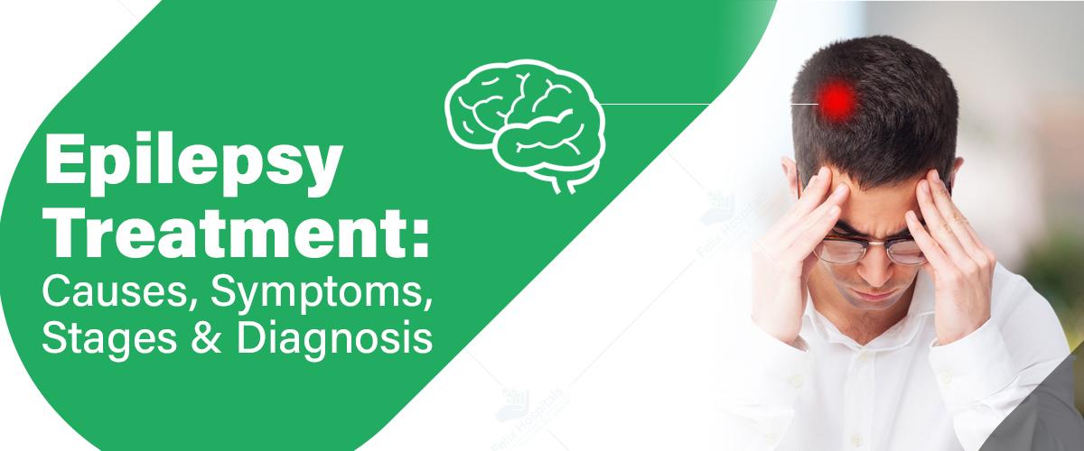
Epilepsy is a neurological disorder in which nerve cells in the brain fail to signal correctly, leading to seizures characterized by uncontrolled bursts of electrical activity that affect sensations, behaviors, awareness, and muscle movements. Although there is no cure for epilepsy, various treatment options are available, and up to 70% of individuals with epilepsy can manage the condition effectively with medications. While finding the best neurology hospital in Noida is essential for addressing urological issues, it is equally important to seek specialized care for neurological conditions like epilepsy to ensure comprehensive management and optimal health outcomes.
Contact us today to schedule a consultation with our expert team and explore the best treatment options tailored to your needs. Call Now - +91 9667064100.
Epilepsy is a chronic condition characterized by recurrent seizures caused by abnormal electrical activity in damaged brain cells. During a seizure, there is a burst of uncontrolled electrical impulses in the brain, which can lead to changes in awareness, muscle control (such as twitching or jerking), sensations, emotions, and behavior. Epilepsy is also known as a seizure disorder.
Epilepsy is a neurological disorder characterized by recurrent seizures, which progress through several stages:
This stage occurs hours or even days before a seizure and is characterized by early warning signs that indicate an impending seizure. Not everyone with epilepsy experiences this stage, but for those who do, it can be a useful time for taking preventive actions or medications. Common symptoms include:
Irritability or mood swings
Headaches
Sleep disturbances
Anxiety or restlessness
Difficulty concentrating
The aura stage is often considered the first part of the actual seizure. It usually lasts a few seconds to a couple of minutes and serves as a warning that a more intense seizure is about to occur. The symptoms of the aura stage depend on which part of the brain is affected and may include:
Visual disturbances (flashing lights, spots)
Unusual smells or tastes
Déjà vu or strange feelings of familiarity
Numbness or tingling in certain body parts
Sudden intense emotions, such as fear or euphoria
Not all patients experience an aura, but when they do, it can help them prepare for an oncoming seizure.
The ictal stage is the actual seizure, and its characteristics vary based on the type of epilepsy. It can last from a few seconds to a few minutes, and the symptoms depend on the region of the brain where the abnormal electrical activity occurs. Seizures can be classified into different types:
Occur in one part of the brain
May involve repetitive movements (e.g., jerking of a limb)
Can cause confusion or a loss of awareness
Affects both sides of the brain
Can cause loss of consciousness or convulsions
Includes subtypes such as tonic-clonic (grand mal) seizures, absence seizures, and myoclonic seizures
Muscle stiffness or rigidity (tonic phase)
Jerking movements (clonic phase)
Loss of consciousness
Sudden collapse
Changes in breathing patterns
Involuntary movements, such as lip-smacking or picking at clothes
This stage occurs immediately after the seizure, and the person may experience several physical and emotional symptoms as the brain recovers from the abnormal electrical activity. The length of this stage varies depending on the type of seizure and the individual’s response. Common symptoms include:
For generalized tonic-clonic seizures, the postictal stage can be longer and more severe compared to focal seizures.
The interictal stage refers to the period between seizures when a person with epilepsy does not experience any symptoms. This stage can last for minutes, hours, days, or even months, depending on the individual’s seizure frequency. During this period, many individuals with epilepsy can function normally, though some may experience ongoing neurological or psychological effects depending on the severity and frequency of their seizures.
In some cases, this stage may involve interictal spikes, where abnormal electrical activity occurs in the brain without triggering a full seizure. This can sometimes be detected through an EEG (electroencephalogram) test.
Epilepsy can result from a variety of factors, which can be broadly categorized into genetic, structural, metabolic, immune, infectious, and unknown causes.
Genetic Factors: Certain types of epilepsy have a genetic component and can run in families. Mutations in specific genes can affect neuronal activity, leading to seizures.
Structural Causes: Brain abnormalities, such as tumors, stroke, congenital malformations, or traumatic brain injuries, can lead to epilepsy.
Metabolic Causes: Disorders that affect the body's metabolism, such as mitochondrial diseases, can cause epilepsy.
Immune Causes: Autoimmune diseases where the body's immune system attacks brain tissues can result in seizures.
Infectious Causes: Infections such as meningitis, encephalitis, or neurocysticercosis can lead to epilepsy.
Unknown Causes: In many cases, the exact cause of epilepsy remains unidentified despite thorough investigations.
The primary symptom of epilepsy is recurrent seizures. However, the manifestation of these seizures can vary widely depending on the type of epilepsy and the areas of the brain involved. Common seizure types include:
Tonic-Clonic Seizures: Characterized by loss of consciousness, muscle stiffening (tonic phase), followed by rhythmic muscle contractions (clonic phase).
Absence Seizures: Brief, sudden lapses in consciousness, often described as staring spells.
Myoclonic Seizures: Sudden, brief jerks or twitches of the muscles.
Atonic Seizures: Sudden loss of muscle tone, causing the person to collapse.
Tonic Seizures: Sudden stiffening of muscles, usually lasting less than 20 seconds.
Clonic Seizures: Repeated jerking movements of muscles on both sides of the body.
Simple Focal Seizures: Affect a small part of the brain and do not impair consciousness. Symptoms can include twitching, unusual sensations, or changes in taste and smell.
Complex Focal Seizures: Affect a larger part of the brain and can alter consciousness, leading to confusion, repetitive movements, or changes in behavior.
Secondary Generalized Seizures: Begin as focal seizures and then spread to both sides of the brain, evolving into generalized seizures.
Diagnosing epilepsy involves a comprehensive evaluation, including:
Medical History: Detailed history of seizures, including the age of onset, frequency, duration, triggers, and family history.
Neurological Examination: Assessment of neurological function, including reflexes, muscle strength, coordination, and sensory abilities.
Electroencephalogram (EEG): A test that measures electrical activity in the brain, helping to identify abnormal patterns associated with epilepsy.
Imaging Studies: MRI or CT scans to detect structural abnormalities in the brain that could be causing seizures.
Blood Tests: To rule out metabolic or genetic conditions that might be contributing to seizures.
While epilepsy is a chronic condition, it can be effectively managed with appropriate treatment. The primary goals of epilepsy treatment are to control seizures, minimize side effects, and improve quality of life. Treatment options include:
Medications: Anti-epileptic drugs (AEDs) are the most common treatment for epilepsy. They help to control seizures by stabilizing electrical activity in the brain. The choice of medication depends on the type of seizures, patient age, overall health, and response to previous treatments.
Surgery: For patients who do not respond to medication, surgical options may be considered. Surgery aims to remove or alter the area of the brain where seizures originate.
Vagus Nerve Stimulation (VNS): A device implanted under the skin sends electrical impulses to the vagus nerve, helping to reduce the frequency and intensity of seizures.
Ketogenic Diet: A high-fat, low-carbohydrate diet that has been shown to reduce seizures in some individuals, particularly children.
Responsive Neurostimulation (RNS): A device implanted in the brain detects abnormal electrical activity and delivers electrical stimulation to prevent seizures.
Lifestyle Modifications: Regular sleep, stress management, avoiding seizure triggers, and maintaining a balanced diet can help manage epilepsy.
Looking for comprehensive care? Explore epilepsy treatment in Noida and regain control of your life.
Living with epilepsy requires ongoing management and support. Individuals with epilepsy should work closely with their healthcare providers to develop a personalized treatment plan. Education and awareness about the condition can help patients and their families better understand and cope with epilepsy. Support groups and counseling services can also provide valuable emotional support and resources.
Dr. Sumit Sharma brings over 10 years of expertise as a neurosurgeon. He specializes in treating a range of neurological and neurosurgical conditions, including brain tumors, brain injuries, spinal tumors, spine injuries, spine fractures, brain and spine tuberculosis, hydrocephalus, migraines, neck pain, back pain, as well as managing depression and anxiety.
Dr. Saumya Mittal brings over 16 years of medical experience to her role, including 2 years as a specialist. Her expertise encompasses a range of neurological conditions, including dementia, seizures, epilepsy, neuropathy, and muscle disorders.
Get in touch now to start your journey towards improved health and well-being. Call Now - +91 9667064100.
Epilepsy, a chronic neurological disorder characterized by recurrent seizures due to abnormal electrical activity in the brain, can be effectively managed with appropriate treatment, although it cannot be cured. Up to 70% of individuals with epilepsy may control their seizures through medications and lifestyle changes. While locating a skilled neurologist in Greater Noida is crucial for addressing urological concerns, seeking specialized care for epilepsy is equally important. If you or a loved one is affected by epilepsy, contact us today to explore the best treatment options and take the first step towards improved management and quality of life.
1.) Is epilepsy a serious disease?
Ans. Yes, epilepsy is considered a serious neurological condition due to the impact of recurrent seizures on daily life. The severity can vary based on the frequency and type of seizures, as well as their effect on a person’s physical and mental health.
2.) What are the main causes of epilepsy?
Ans. The main causes of epilepsy can be categorized into genetic factors, structural causes (like brain tumors or injuries), metabolic disorders, immune causes, infectious diseases, and sometimes the exact cause remains unknown.
3.) Can epilepsy live a normal life?
Ans. Yes, many people with epilepsy can lead normal, fulfilling lives with proper management. Medications, lifestyle changes, and support can help control seizures and minimize their impact.
4.) How does epilepsy affect you physically?
Ans. Physically, epilepsy can lead to symptoms such as muscle jerks or stiffness during seizures, sudden loss of muscle control, or even injuries from falls. The physical effects can vary based on the type of seizures experienced.
5.) Is epilepsy 100% curable?
Ans. Currently, epilepsy is not considered 100% curable. However, many people can achieve significant control over their seizures with medication, lifestyle changes, or other treatments.
6.) Can epilepsy go away naturally?
Ans. In some cases, epilepsy may remit or improve significantly over time, especially in children. However, it does not typically "go away" completely without intervention.
7.) At what age does epilepsy start?
Ans. Epilepsy can start at any age, but it commonly begins in childhood or early adulthood. It can also develop later in life due to various causes, such as brain injury or stroke.
8. Is epilepsy life-ending?
Ans. Epilepsy itself is not usually life-ending, but severe or uncontrolled seizures can lead to complications or increase the risk of injury. With proper management, many people with epilepsy can live a full and healthy life.
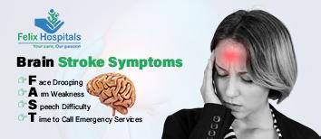
Recognizing the symptoms of a brain stroke is critical for getting prompt medical attention. The acronym F.A.S.T. is often used to remember the most common Brain stroke symptoms.
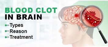
Blood clot in the brain is a serious condition causing sudden neurological symptoms and requires immediate medical treatment.

A concise guide explaining brain tumor surgery cost in Noida, including key factors, treatment options, and expert care.

Multiple sclerosis is a neurological disease that affects the brain and spinal cord.

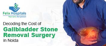
Gallstones are a significant medical condition affecting millions of people worldwide. It is crucial to address and treat gallstones to prevent serious complications such as inflammation, infection, or bile duct blockages. Recognizing and managing symptoms like abdominal pain and digestive issues can help in early intervention. Additionally, seeking care at a specialized gallbladder stone removal hospital ensures proper treatment, alleviating symptoms and reducing the risk of more severe health problems.
Experiencing discomfort from gallstones? Call +91 9667064100 or simply click here for expert advice and relief!
Gallstones are hardened deposits of digestive fluid that form in your gallbladder, a small, pear-shaped organ located on the right side of your abdomen, just beneath your liver. The gallbladder's primary function is to store bile, a digestive fluid produced by the liver, and release it into the small intestine to aid in digestion.
These stones can vary in size, ranging from as small as a grain of sand to as large as a golf ball. Some people may develop a single gallstone, while others may have multiple stones simultaneously.
There are two main types of gallstones:
1. Cholesterol Gallstones: These are the most prevalent type and typically appear yellow. They are primarily composed of undissolved cholesterol but may also contain other substances. Cholesterol gallstones form when there is too much cholesterol in the bile, which the body struggles to break down effectively.
2. Pigment Gallstones: These stones are usually dark brown or black. They develop when bile contains an excess of bilirubin, a substance produced during the breakdown of red blood cells. High levels of bilirubin can lead to the formation of these darker stones, which are less common than cholesterol gallstones.
Many people with gallstones may not experience any symptoms at all. However, if a gallstone lodges in a duct and causes a blockage, you may experience the following signs and symptoms:
Gallstone pain, commonly known as a gallbladder attack, can vary in duration, typically lasting from several minutes to a few hours. This pain, often sharp and intense, occurs when a gallstone temporarily blocks the bile ducts, causing significant discomfort in the upper abdomen.
The exact cause of gallstone formation isn't entirely clear, but medical professionals believe they may result from the following factors:
Excess cholesterol in bile: If your liver produces more cholesterol than your bile can dissolve, the excess cholesterol may form into crystals and eventually into stones.
Excess bilirubin in bile: Certain conditions can cause your liver to produce too much bilirubin, contributing to gallstone formation.
Incomplete gallbladder emptying: If your gallbladder doesn't empty completely or often enough, bile may become very concentrated, leading to stone formation.
Gallstones are a prevalent condition influenced by several risk factors. Recognizing these factors can help in assessing your risk and taking preventive measures. Here are some key factors that may increase your likelihood of developing gallstones:
If gallstones are not addressed promptly, they can lead to a range of serious complications. Understanding these potential issues is crucial for managing the condition effectively and preventing further health problems.
Here are some complications that may arise if gallstones are left untreated:
Inflammation of the gallbladder (cholecystitis): This condition can cause intense pain and fever, leading to significant discomfort.
Blockage of the common bile duct: This may result in severe pain, jaundice, and potential bile duct infection.
Blockage of the pancreatic duct: Such blockage can cause inflammation of the pancreas (pancreatitis), leading to severe abdominal pain and often necessitating hospitalization.
Increased risk of gallbladder cancer: Although rare, a history of gallstones slightly elevates the risk of developing gallbladder cancer.
Gallstones are a prevalent condition that can vary from being asymptomatic to causing severe discomfort and complications. Accurate diagnosis is crucial to determine the appropriate treatment and manage symptoms effectively.
Diagnosis:
Medical History and Physical Examination: The initial assessment often includes reviewing symptoms and medical history, followed by a physical exam to check for abdominal tenderness or pain.
Ultrasound: The most common and effective test for detecting gallstones, using sound waves to create images of the gallbladder and identify stones.
CT Scan: Provides detailed cross-sectional images and is used if the ultrasound results are inconclusive or if complications are suspected.
HIDA Scan: A nuclear imaging test that evaluates the function of the gallbladder and can help detect blockages in the bile ducts.
Endoscopic Retrograde Cholangiopancreatography (ERCP): A procedure combining endoscopy and X-rays to visualize and remove stones from the bile ducts.
Treatment:
Medication: Oral medications like ursodeoxycholic acid can help dissolve cholesterol gallstones over time, though this method is less common due to its slow effectiveness.
Extracorporeal Shock Wave Lithotripsy (ESWL): Uses shock waves to break up gallstones into smaller pieces that can then be passed more easily, often used in conjunction with other treatments.
Cholecystectomy: The surgical removal of the gallbladder, typically performed laparoscopically (minimally invasive) or through open surgery, depending on the case and the presence of complications.
Lifestyle Modifications: Dietary changes such as reducing fat intake and increasing fiber, along with regular exercise, can help manage and prevent gallstone formation.
When considering treatment options for gallstones, it's essential to evaluate the cost of gallbladder surgery removal, as this can vary based on the type of surgery and medical facility. Consulting with a healthcare provider can help you understand the financial aspects and insurance coverage related to the procedure, ensuring you make an informed decision about your treatment plan.
Reducing your risk of developing gallstones involves adopting healthy lifestyle habits. Implementing these strategies can help lower your chances of experiencing this condition.
Here are some effective tips to consider:
Maintain regular meal times: Consistent meal schedules are important; avoid skipping meals or fasting as these practices can increase the risk of gallstones.
Lose weight gradually: If weight loss is needed, aim for a gradual reduction of 1-2 pounds per week to avoid rapid weight loss, which can trigger gallstones.
Increase fiber intake: Enhance your diet with fiber-rich foods such as fruits, vegetables, and whole grains to support digestive health and reduce gallstone risk.
Maintain a healthy weight: Obesity and being overweight are significant risk factors. Focus on achieving and sustaining a healthy weight through a balanced diet and regular physical activity.
If you notice any concerning signs or symptoms, it is crucial to consult your doctor promptly. Seek immediate medical attention if you experience signs of a severe gallstone complication, including:
Dr. Ritesh Agarwal
Dr. Ritesh Agarwal, with 15 years of experience in general, minimal access, and laparoscopic surgery, is highly skilled in treating gallstones. His expertise encompasses a wide range of procedures, including laparoscopic cholecystectomy (gallbladder stone removal), hernia repairs, and various proctology surgeries, ensuring comprehensive care for his patients.
Gallstones are a common medical condition that can range from asymptomatic to severely painful. They can lead to serious complications if left untreated, impacting your overall health and quality of life. By understanding the risk factors and adopting preventive measures, you can reduce your chances of developing gallstones. Regular check-ups and a proactive approach to managing your health are essential. If you suspect you have gallstones or experience any concerning symptoms, don't hesitate to consult with a gallbladder specialist for proper diagnosis and treatment.
Struggling with gallstone symptoms? Reach out to +91 9667064100 for professional guidance and support today!
Is it acceptable to leave gallstones untreated?
Leaving gallstones untreated may be acceptable if they are asymptomatic and not causing any problems. However, if they lead to pain or complications, treatment or removal is often necessary to prevent further issues.
Does a 1 cm gallstone require surgery?
A 1 cm gallstone may require surgery if it causes symptoms or complications. The decision to proceed with surgery depends on factors like pain, risk of blockage, and the overall health of the patient.
Can gallstones damage the liver?
Gallstones can potentially lead to liver damage if they cause blockages in the bile ducts, leading to infections or inflammation. These blockages can affect liver function and contribute to complications if not treated.
How long is the recovery period after gallbladder surgery?
Recovery time after gallbladder surgery typically involves a short period of bed rest. Most people can return to normal activities within a week, although full recovery may take several weeks depending on the type of surgery and individual health.
Is it possible to live without a gallbladder?
Yes, it is possible to live without a gallbladder. The body can still digest food, though some people may need to adjust their diet and may experience changes in digestion.
Why remove the gallbladder rather than just the stones?
Removing the gallbladder is often preferred because it addresses the root cause of gallstones and prevents future occurrences. Stones can recur if the gallbladder remains, leading to repeated issues and potential complications.
What foods should be avoided after gallbladder removal?
After gallbladder removal, it is advisable to avoid high-fat and greasy foods, as well as large meals. These can be harder to digest without the gallbladder and may cause discomfort or digestive issues.
How long does gallbladder surgery typically take?
Gallbladder surgery usually takes between 1 to 2 hours, depending on whether it is performed laparoscopically or as an open procedure. The duration can vary based on the complexity of the case and the surgeon’s expertise.

Understand the factors that influence gallbladder stone surgery cost in Noida, including hospital charges, surgeon expertise, type of procedure, and post-operative care expenses.
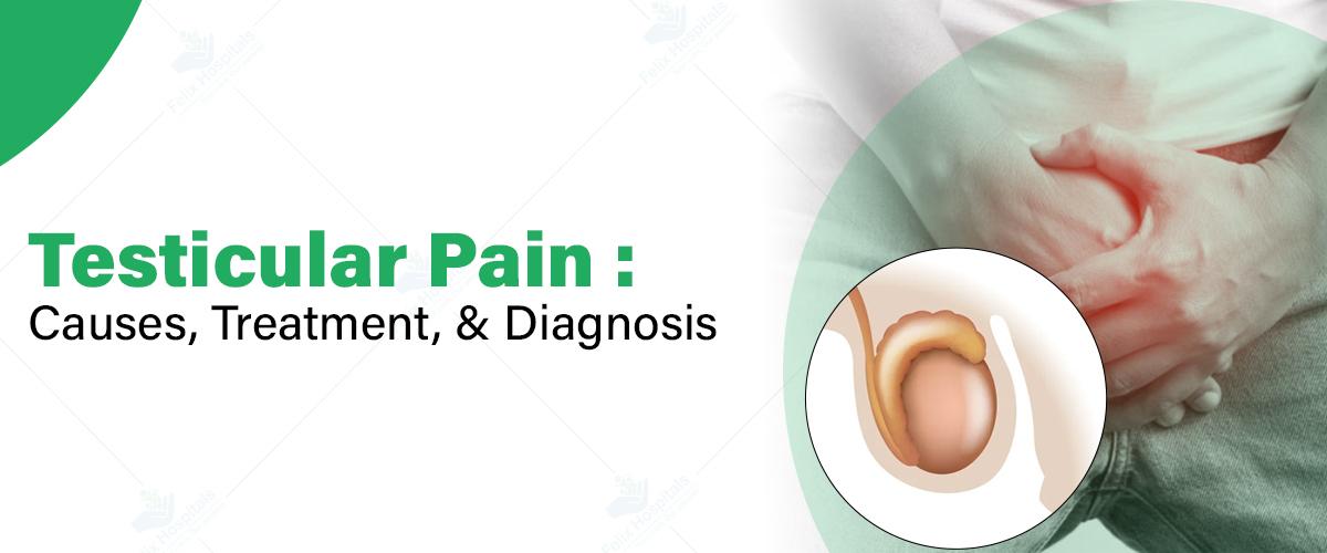
Testicular pain is a condition that can cause significant discomfort and concern for many men. Although it’s a sensitive topic, understanding its causes, treatment options, and diagnostic processes is crucial for maintaining male reproductive health. Let’s explore the factors behind testicular pain to help you understand when to seek medical help and how to manage the condition effectively. But first, let’s understand what causes this pain and how it can impact your health.
Testicular pain refers to discomfort or pain experienced in one or both testicles. The severity can vary from mild to severe and may be accompanied by symptoms such as swelling, redness, or changes in the appearance of the scrotum. Persistent or severe pain should be evaluated by a healthcare professional, as some underlying causes can be serious and require prompt treatment.
Contact us at +91 9667064100 to schedule your appointment at Noida's top hospital for effective treatment of testicular pain.
Testicular pain can originate from various sources, ranging from minor injuries to more serious health conditions. Understanding these causes is crucial for accurate diagnosis and effective treatment. Here’s a breakdown of some common causes:
1. Trauma or Injury
Physical trauma to the testicles or scrotum is a frequent cause of acute pain. This can occur due to:
Even minor impacts can result in significant discomfort due to the sensitivity of the testicular area.
2. Epididymitis
Epididymitis involves inflammation of the epididymis, the coiled tube at the back of the testicle that stores and carries sperm. It is often caused by bacterial infections and may present with:
3. Orchitis
Orchitis is the inflammation of one or both testicles, commonly due to viral or bacterial infections. It can occur alongside epididymitis (epididymo-orchitis) and may lead to:
4. Testicular Torsion
Testicular torsion is a medical emergency where the spermatic cord becomes twisted, cutting off blood flow to the testicle. It’s symptoms may include:
Immediate medical intervention is necessary to prevent loss of the testicle.
5. Varicocele
A varicocele Treatment In Noida is an enlargement of the veins that drain the testicle, similar to varicose veins. It can cause:
6. Inguinal Hernia
An inguinal hernia occurs when part of the intestine protrudes through a weak spot in the abdominal wall into the scrotum. This can lead to:
7. Kidney Stones
Although kidney stones are not directly related to the testicles, they can cause referred pain in the testicular area. This pain is often accompanied by:
8. Infections
Various infections can lead to testicular pain, including:
9. Testicular Cancer
Though rare, testicular cancer is a serious condition, particularly in younger men. It may present as:
Accurately diagnosing testicular pain is crucial for effective treatment, and seeking care at the best urology hospital in Noida can ensure a comprehensive evaluation. The diagnostic process generally involves several key steps to identify the underlying cause and guide appropriate treatment.
1. Medical History
To start, your doctor will gather a detailed medical history. This includes questions about the nature, onset, and duration of the pain, any recent injuries or infections, your sexual history, and other associated symptoms. Understanding these factors helps in identifying the underlying cause of the pain.
2. Physical Examination
A thorough physical examination follows, where the doctor inspects the scrotum for any signs of swelling, redness, or lumps. They will palpate the testicles and surrounding areas to check for tenderness or abnormalities and assess the abdomen and groin for any additional issues that might be contributing to the pain.
3. Urinalysis
A urinalysis is conducted to detect potential issues in the urinary system. This test can reveal signs of infection, the presence of blood, and other abnormalities in the urine that could be related to the testicular pain.
4. Blood Tests
Blood tests are often performed to look for indicators of infection, such as an elevated white blood cell count, and to check for tumor markers if there is a suspicion of testicular cancer. These tests provide important information that aids in diagnosing the cause of the pain.
5. Imaging Studies
Imaging studies are used to get a clearer view of the internal structures. An ultrasound is the primary method for obtaining detailed images of the testicles and surrounding areas. A CT scan may be used to examine the abdomen or pelvis if related issues are suspected. Occasionally, an MRI is used for more detailed imaging when necessary.
6. Surgical Exploration
In emergencies, such as suspected testicular torsion, immediate surgical exploration might be required. This procedure allows for both diagnosis and treatment, helping to address any urgent issues that could lead to serious complications if not managed promptly.
Testicular pain can arise from various causes, ranging from minor issues to serious medical conditions. Effective treatment depends on accurately diagnosing the underlying cause. Here’s an overview of the common treatment approaches for managing testicular pain, tailored to different causes and conditions.
1. Pain Management
To manage testicular pain, use over-the-counter pain relievers like ibuprofen or acetaminophen. Apply cold or warm compresses for temporary relief and wear supportive underwear to reduce discomfort and provide stability.
2. Antibiotics
For bacterial infections such as epididymitis or orchitis, antibiotics are prescribed to eliminate the infection and relieve pain. Adhering to the prescribed regimen is crucial for effective treatment.
3. Antiviral Medications
In cases of viral infections like mumps-related orchitis, antiviral medications help manage symptoms and reduce inflammation, aiding in recovery and pain relief.
4. Surgery
Surgery may be needed for conditions like testicular torsion, large hydroceles, or severe varicoceles. Emergency surgery is critical for testicular torsion to restore blood flow and prevent complications.
5. Cancer Treatment
If diagnosed with testicular cancer, treatment may include surgery (orchiectomy), chemotherapy, and radiation therapy to remove cancerous cells and manage pain.
6. Lifestyle Modifications
To manage symptoms, wear supportive underwear, avoid heavy lifting, and practice safe sex. These adjustments can help alleviate pain and prevent further issues.
7. Follow-up Care
Regular follow-up appointments are essential to monitor progress, adjust treatments, and address any new or persistent symptoms, ensuring effective management of the condition.
While some causes of testicular pain may resolve independently, you should seek medical help if you experience:
Although not all causes of testicular pain are preventable, you can reduce the risk by:
Explore the expertise of best urologists in Noida Renowned for their advanced skills in diagnosing and treating testicular pain. These specialists offer comprehensive care tailored to the specific needs of each patient.
Dr. Bhanwar Lal Barkesiya, MBBS, MS, FMAS, and MCH (Urology) (Gold Medalist), boasts over 9 years of specialized experience in managing testicular pain and related conditions. His proficiency includes a range of procedures such as URSL, PCNL, and ureterolithotomy, as well as advanced techniques like robotic-assisted surgeries. Dr. Barkesiya is known for his thorough approach to diagnosing and treating various urological issues, ensuring exceptional care.
Dr. Sachin Khandelwal, MBBS, MS (Gen Surg), MS (Urology), is a distinguished urologist with more than 12 years of experience in addressing testicular pain and other urological conditions. His expertise extends to managing kidney blockages, bladder issues, urinary tract infections, and performing both laparoscopic and open surgeries. Dr. Khandelwal is also well-versed in renal transplantation and endourology, providing comprehensive solutions for complex urological concerns.
Experiencing testicular pain and seeking top-notch care? Call +91 9667064100 to consult with the leading urologists in Noida.
Testicular pain can result from a range of issues, from minor injuries to serious conditions. Understanding the symptoms, causes, and available treatments is key to managing testicular health effectively. Although discussing testicular pain may be uncomfortable, prompt medical attention is crucial for preventing complications and ensuring optimal outcomes. By staying informed and proactive, men can address testicular pain, maintain reproductive health, and achieve peace of mind.
1.) Can testicular pain be a sign of a serious condition?
Yes, testicular pain can indicate serious conditions like testicular torsion, infections, or tumors. It's crucial to consult a healthcare provider for a proper diagnosis and treatment to rule out any severe underlying issues.
2.) Are there any at-home remedies for relieving testicular pain?
At-home remedies include applying ice packs, wearing supportive underwear, and avoiding activities that aggravate the pain. However, these methods are not a substitute for medical evaluation, especially if the pain persists or worsens.
3.) Can testicular pain affect fertility?
Testicular pain itself might not directly affect fertility, but underlying conditions causing the pain, such as infections or varicoceles, can potentially impact sperm production or function. Addressing the root cause with a healthcare provider is essential for preserving fertility.
4.) Are there any lifestyle changes that can help manage testicular pain?
Lifestyle changes include wearing supportive underwear, avoiding prolonged sitting, and managing stress. Regular exercise and maintaining a healthy weight can also help. However, these adjustments should complement, not replace, professional medical treatment.
5.) Can testicular pain be a symptom of a hernia?
Yes, testicular pain can be a symptom of an inguinal hernia, where abdominal tissue pushes into the groin area. This condition can cause discomfort in the testicles and requires medical evaluation for appropriate treatment and management.

In the realm of modern medicine, radiodiagnosis plays a pivotal role in the accurate detection, diagnosis, and monitoring of various medical conditions. As medical technology continues to advance, the importance of high-quality radiodiagnosis services has never been greater. This guide explores the key aspects of top-quality radiodiagnosis services, highlighting what patients should look for to ensure they receive the best possible care at the Best Radiodiagnosis Services Hospital.
Schedule your consultation today to benefit from advanced imaging technologies and expert interpretations that ensure accurate and timely diagnosis. Call Now - +91 9667064100.
Radiodiagnosis, often referred to as diagnostic imaging, encompasses a range of techniques that utilize various forms of energy, such as X-rays, sound waves, magnetic fields, and radioactive substances, to create images of the body’s internal structures. These images are crucial for diagnosing diseases, guiding treatment plans, and monitoring the progress of medical interventions.
Radiodiagnosis services are essential for early detection of conditions such as cancer, cardiovascular diseases, neurological disorders, and musculoskeletal problems. By providing detailed and precise images, these services enable healthcare providers to make informed decisions, leading to better patient outcomes.
1. Advanced Technology: The cornerstone of any top-tier radiodiagnosis service is the use of state-of-the-art imaging technology. This includes high-resolution CT (Computed Tomography) scanners, MRI (Magnetic Resonance Imaging) machines, digital X-rays, and ultrasound equipment. The latest advancements in these technologies allow for faster, clearer, and more accurate imaging, reducing the need for repeat scans and ensuring timely diagnosis.
2. Expert Radiologists: A critical factor in the quality of radiodiagnosis services is the expertise of the radiologists interpreting the images. Radiologists are medical doctors specialized in diagnosing and treating conditions using medical imaging. The best services are staffed by experienced radiologists who are not only skilled in interpreting a wide range of imaging modalities but also collaborate closely with other medical specialists to provide comprehensive patient care.
3. Comprehensive Imaging Services: Leading radio diagnosis centers offer a full spectrum of imaging services, ensuring that patients have access to the most appropriate diagnostic tools for their specific needs. These services typically include:
4. Patient-Centered Care: High-quality Radiodiagnosis Services prioritize patient comfort and convenience. This includes offering flexible scheduling, minimizing wait times, and providing clear communication throughout the imaging process. Additionally, the best centers employ advanced techniques that reduce patient exposure to radiation while maintaining image quality.
5. Accurate and Timely Reporting: The speed and accuracy of image interpretation are critical in ensuring that patients receive timely diagnosis and appropriate treatment. Leading radiodiagnosis services utilize digital platforms that allow for rapid image processing and sharing, enabling healthcare providers to make swift and informed decisions.
6. Collaborative Approach: Top radiodiagnosis services emphasize a collaborative approach to patient care. Radiologists work closely with referring physicians, surgeons, and other healthcare professionals to ensure that imaging results are seamlessly integrated into the overall treatment plan. This multidisciplinary approach not only enhances the effectiveness of patient care but also supports better health outcomes while considering factors such as the radiodiagnosis services treatment cost, ensuring that patients receive comprehensive and cost-effective care.
When selecting a radio diagnosis service, patients should consider several factors:
Dr. Pulkit Soni is a distinguished Radiologist with over 5 years of experience, bringing a robust clinical background and advanced expertise in Radiodiagnosis. His proficiency in utilizing Healthcare Artificial Intelligence significantly enhances diagnostic accuracy and improves patient outcomes, making him a key asset to our team.
Visit our website [email protected] for more information on our services and how we can support your health journey.
Radiodiagnosis services are a cornerstone of modern healthcare, providing essential insights that guide the diagnosis and treatment of a wide range of medical conditions. By choosing a radiodiagnosis service that offers advanced technology, expert radiologists in Noida, comprehensive imaging options, and a patient-centered approach, individuals can ensure they receive the best possible diagnostic care. As medical technology continues to evolve, the role of high-quality Radiodiagnosis Services will only become more vital in supporting effective and timely healthcare.
1. What are the different types of radiodiagnosis services?
Radiodiagnosis services encompass a variety of imaging techniques used to visualize the internal structures of the body. Key types include X-rays, computed tomography (CT) scans, magnetic resonance imaging (MRI), ultrasound, and nuclear medicine scans such as positron emission tomography (PET) scans and single-photon emission computed tomography (SPECT).
2. What does radiodiagnosis involve?
Radiodiagnosis involves using imaging technologies to capture detailed images of the body's internal structures. These images help diagnose diseases, monitor the progression of conditions, and evaluate the effectiveness of treatments. The process typically includes capturing the images, interpreting them, and integrating the findings with clinical information to guide patient care.
3. How does radiology differ from radiodiagnosis?
Radiology is the broader medical field that encompasses the use of imaging technologies to diagnose and treat diseases. Radiodiagnosis, a subset of radiology, specifically focuses on the diagnostic aspect of imaging. While radiology includes both diagnostic and interventional procedures, radiodiagnosis is concerned primarily with interpreting images to diagnose medical conditions.
4. What is the role of radiodiagnosis in healthcare?
Radiodiagnosis plays a crucial role in healthcare by providing essential information for diagnosing diseases, planning treatments, and monitoring the effectiveness of therapies. It helps in early detection of conditions, accurate assessment of disease extent, and guiding therapeutic interventions, ultimately contributing to improved patient outcomes.
5. What functions does diagnostic radiology serve?
Diagnostic radiology serves several functions, including detecting and diagnosing diseases, evaluating the severity and progression of conditions, guiding treatment plans, monitoring patient responses to treatments, and aiding in surgical planning. It provides critical insights that assist healthcare providers in making informed medical decisions.
6. What is the purpose of radiology treatment?
The purpose of radiology treatment is to utilize imaging techniques to identify and monitor medical conditions. This includes diagnosing diseases, evaluating the extent of injuries or illnesses, and assessing the effectiveness of ongoing treatments. Radiology treatments aim to improve patient care by providing detailed and accurate internal views of the body.
7. Why do patients undergo radiological examinations?
Patients undergo radiological examinations to obtain detailed images of their internal organs and tissues. These images help in diagnosing medical conditions, assessing the severity of diseases, planning and monitoring treatments, and guiding surgical procedures. Radiological examinations are essential for accurate diagnosis and effective management of various health issues.

Radiology is a vital medical field that helps doctors diagnose and treat diseases using advanced imaging techniques like X-rays, CT scans, MRI, and ultrasound.

Interventional radiology (IR) is a medical specialty that uses imaging guidance (such as X-ray, CT scan, ultrasound, or MRI).
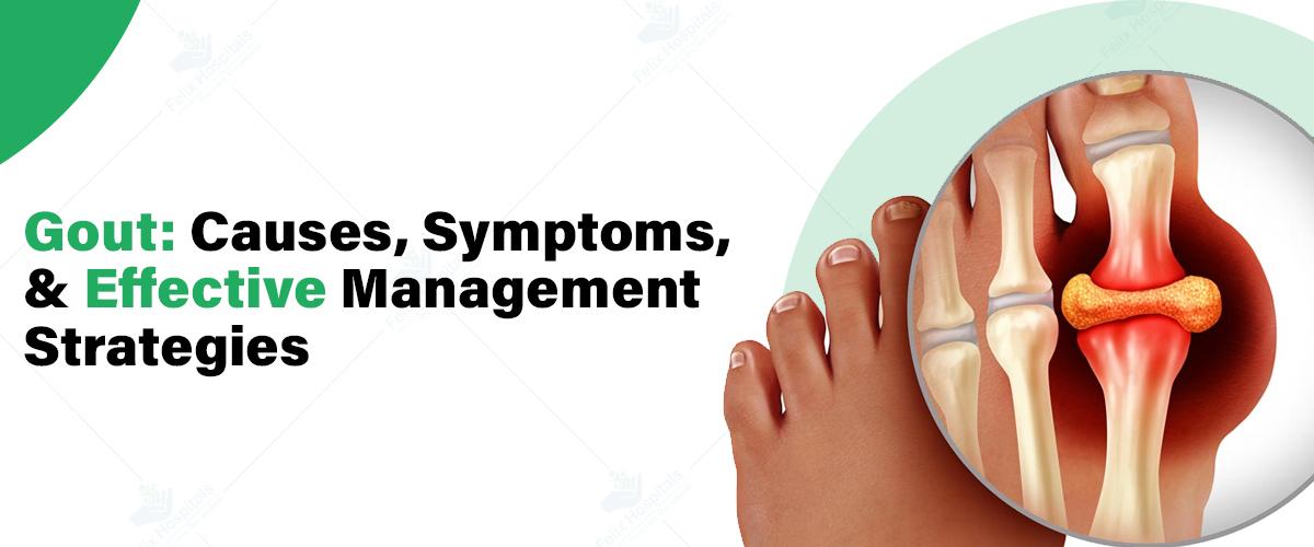
Gout is a form of inflammatory arthritis that has troubled individuals for centuries, marked by sudden, severe joint pain, swelling, and redness, typically affecting the big toe. Once thought to be a disease of the affluent due to its link with a rich diet, gout now affects people from all backgrounds.
For those seeking comprehensive management and treatment, consulting specialists at the best rheumatology hospital can provide valuable insights and effective strategies to control this debilitating condition. Let’s explore the causes, symptoms, and management techniques for gout, empowering you with the knowledge to take control of your health.
Click here or call +91 9667064100 and get expert help with your gout for a pain-free tomorrow!
Gout is caused by the buildup of uric acid crystals in the joints, which triggers an inflammatory response. Uric acid is a byproduct of the breakdown of purines, which are found in various foods and are also produced naturally by the body.
When the body produces too much uric acid or is unable to efficiently remove it, the excess uric acid can crystallize and accumulate in the joints, leading to the development of gout.
Gout is caused by the accumulation of urate crystals in the joints, which leads to intense pain and inflammation. This happens when there are high levels of uric acid in the blood, a condition often linked to the following factors:
Purine Metabolism: Uric acid is produced when your body breaks down purine which is a substance found both naturally in your body and in certain foods. High purine levels from red meat, and some seafood can lead to elevated uric acid levels.
Diet: Foods rich in purines and beverages like beer and sugary drinks can increase uric acid levels. Consuming these in excess contributes to higher uric acid concentrations in the blood.
Kidney Function: Normally, uric acid dissolves in the blood and is excreted through the kidneys into the urine. However, if the kidneys do not excrete enough uric acid or if the body produces too much, uric acid can accumulate.
Genetics and Health Conditions: Genetic factors and certain health conditions, such as obesity, high blood pressure, and kidney disease, can also play a role in increasing uric acid levels or affecting its elimination.
Dehydration: Inadequate fluid intake can lead to higher uric acid concentrations, as it reduces the amount of uric acid that is excreted through the urine.
When uric acid levels are too high, urate crystals can form in the joints or surrounding tissues, causing the characteristic pain, inflammation, and swelling associated with gout.
The hallmark symptom of gout is a sudden, severe, and often excruciating joint pain, typically affecting the big toe. However, gout can also occur in other joints, such as the ankles, heels, knees, and wrists.
Other common symptoms of gout include:
Intense joint swelling and inflammation
Redness and warmth in the affected joint
Decreased range of motion and difficulty moving the joint
Fever and chills in some cases
Gout attacks can occur suddenly, often waking individuals from sleep, and can last for several days to weeks if left untreated.
Diagnosing gout involves several key steps to ensure an accurate assessment:
Medical History and Physical Examination: Your healthcare provider will begin by discussing your symptoms, reviewing your medical history, and inquiring about your dietary habits. This helps to understand the context of your condition and any potential contributing factors.
Joint Fluid Analysis: To confirm gout, a small sample of fluid may be extracted from the affected joint using a needle. This fluid is then examined under a microscope to identify the presence of uric acid crystals, which are indicative of gout.
Blood Tests: A blood test measures the level of uric acid in your bloodstream. Elevated levels of uric acid can support the diagnosis of gout, though it’s important to note that high uric acid levels alone do not confirm gout, as they can be present without symptoms.
Imaging Tests: X-rays, ultrasound, or other imaging techniques may be used to rule out other potential causes of joint pain and inflammation. These tests can help visualize the extent of joint damage and support the gout diagnosis by showing characteristic changes in the joint.
An accurate diagnosis is essential because gout can be mistaken for other forms of arthritis or joint disorders. Proper diagnosis ensures that the appropriate treatment plan is developed for effective management of the condition.
Effective management of gout involves a combination of lifestyle modifications, medication, and ongoing monitoring. The primary goals of gout management are to:
Relieve the acute symptoms of a gout attack
Prevent future gout attacks
Reduce the risk of complications, such as joint damage or kidney stones
Gout can be managed through the following approaches:
Dietary changes: Limiting the intake of purine-rich foods, such as red meat, seafood, and sugary beverages, can help lower uric acid levels.
Hydration: Drinking plenty of water and other fluids can help flush out excess uric acid from the body.
Weight management: Maintaining a healthy weight can reduce the risk of gout by lowering uric acid levels.
Exercise: Regular physical activity can help improve overall health and reduce the risk of gout.
Anti-inflammatory drugs: Nonsteroidal anti-inflammatory drugs (NSAIDs), such as ibuprofen or naproxen, can help reduce inflammation and relieve the pain of a gout attack.
Corticosteroids: Oral or injectable corticosteroids may be used to quickly reduce inflammation and pain during a gout flare-up.
Uric acid-lowering medications: Drugs like allopurinol or febuxostat can help lower uric acid levels and prevent future gout attacks.
Uricosuric medications: These medications, such as probenecid, work by increasing the excretion of uric acid from the body.
It is important to work closely with your healthcare provider to determine the most appropriate medication regimen for your individual needs and to monitor for any side effects.
Regular check-ups with your healthcare provider are crucial for managing gout effectively.
This may include:
Periodic blood tests to monitor uric acid levels
Discussions about dietary and lifestyle modifications
Adjustments to medication dosages or regimens as needed
Monitoring for any potential complications, such as kidney stones or joint damage
By taking a proactive and comprehensive approach to gout management, individuals can significantly reduce the frequency and severity of gout attacks, minimize the risk of complications, and improve their overall quality of life.
In addition to the management strategies mentioned above, there are several steps individuals can take to help prevent future gout attacks:
Maintain a healthy body weight: Excess weight, especially around the midsection, can increase the risk of gout.
Limit intake of high-purine foods: Avoid or limit consumption of red meat, seafood, and sugary beverages.
Stay hydrated: Drink plenty of water and other fluids throughout the day.
Manage underlying medical conditions: Conditions like diabetes, high blood pressure, and kidney disease can increase the risk of gout, so it's important to keep them under control.
Avoid sudden changes in diet or alcohol consumption: Drastic changes can trigger a gout attack.
By implementing these preventive measures, individuals with gout can take an active role in managing their condition and reducing the frequency and severity of future attacks.
Dr. Kiran Seth is a highly experienced Rheumatologist with over 15 years of expertise in treating and managing various rheumatic conditions, including Gout. She specializes in Immunotherapy, using biologics for complex autoimmune diseases like Rheumatoid Arthritis and Systemic Lupus Erythematosus (SLE). Dr. Seth is well-known for her skill in treating musculoskeletal diseases and other conditions such as Osteoarthritis, Osteoporosis, and Ankylosing Spondylitis. Her comprehensive approach and patient-centered care make her a trusted specialist in the field of Rheumatology.
Gout is a complex and often debilitating condition, but with proper management and a proactive approach, individuals can take control of their symptoms and reduce the risk of complications. By understanding the underlying causes, recognizing the symptoms, and implementing effective management strategies, those living with gout can enjoy a better quality of life and minimize the impact of this chronic condition.
If you or a loved one are experiencing symptoms of gout, it’s essential to consult with a healthcare provider or the Best Rheumatologists in Noida who can provide an accurate diagnosis and develop a personalized treatment plan. With the right support and guidance, individuals with gout can live fuller, more active lives.
Struggling with gout and need professional help? Contact us at +91 9667064100 for the best gout treatment in Noida.
Q1. Are gout and arthritis the same?
Ans. No, gout and arthritis are not the same. Gout is a specific type of arthritis caused by uric acid crystals in the joints, while arthritis is a general term for inflammation of the joints that includes various conditions such as osteoarthritis and rheumatoid arthritis.
Q2. Is gout a lifelong condition?
Ans. Gout can be a lifelong condition, but it can be managed effectively with treatment. With proper medication and lifestyle changes, individuals can control symptoms, prevent flare-ups, and maintain a good quality of life.
Q3. What foods should you avoid if you have gout?
Ans. If you have gout, avoid foods high in purines, such as red meat, organ meats, shellfish, and sugary beverages. Also, limit alcohol and foods rich in fructose, as these can increase uric acid levels and trigger gout attacks.
Q4. How is gout diagnosed?
Ans. Gout is diagnosed through a combination of methods, including a physical examination, blood tests to check uric acid levels, and analysis of joint fluid for uric acid crystals. Sometimes, imaging tests like X-rays or ultrasound may be used to assess joint damage.
Q5. What are the four stages of gout?
Ans. The four stages of gout are: 1) Asymptomatic hyperuricemia, where high uric acid levels occur without symptoms; 2) Acute gout, characterized by sudden and severe pain; 3) Intercritical gout, a symptom-free period between attacks; and 4) Chronic tophaceous gout, where persistent symptoms and joint damage occur.
Q6. Can drinking water help flush out uric acid?
Ans. Yes, drinking plenty of water can help flush out uric acid from the body. Staying hydrated supports kidney function, which aids in the excretion of uric acid, potentially reducing the risk of gout attacks.
Q7. How can you lower uric acid in three days?
Ans. To lower uric acid levels quickly, stay well-hydrated, avoid high-purine foods, and consider taking prescribed medications or over-the-counter remedies as directed by your healthcare provider. A temporary reduction in uric acid levels can help manage symptoms more effectively.

Knock knees, medically known as genu valgum, is a common condition in young children where the knees angle inward and touch one another when the legs are straightened.
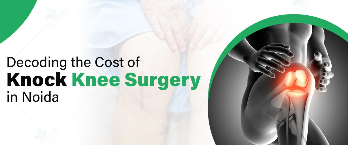
Knock knee surgery, or genu valgum correction, is a surgical procedure aimed at correcting the alignment of the knees in individuals with knock knees. This condition, where the knees angle inward and touch each other when the legs are straightened, can lead to pain, difficulty walking, and other complications. In this blog, we will explore the cost of knock knee surgery, highlight factors affecting the cost, and offer tips to manage and potentially reduce expenses.
Call +91 9667064100 now for a quick cost estimate on knock knee surgery.
Knock knee surgery is designed to correct the alignment of the knees by adjusting the bone structure. This can involve various surgical techniques, such as osteotomy or guided growth surgery, to realign the bones and improve function. The goal is to alleviate symptoms, improve mobility, and enhance overall quality of life. It's crucial to choose a skilled orthopedic surgeon and facility for optimal results.
The cost of knock knee surgery in Noida can vary based on the complexity of the procedure, hospital, and other factors. Here’s a breakdown of typical costs:
| Minimum Cost | ₹1,00,000 approx |
| Average Cost | ₹2,20,000 approx |
| Maximum Cost | ₹1,40,000 approx |
Felix Hospital offers the most cost-effective and economically viable options for undergoing knock knee surgery in Noida.
| Minimum Cost | ₹ 70,000 approx |
| Average Cost | ₹ 2, 00,000 approx |
| Maximum Cost | ₹1,40,000 approx |
Several factors can influence the total cost of knock knee surgery:
Hospital and Surgeon Fees
Hospital Choice: Select an arthritis hospital with a reputation for quality care and reasonable pricing.
Surgeon’s Expertise: Experienced surgeons may charge higher fees but often provide better outcomes.
Pre-Operative Costs
Consultations: Initial consultations with orthopedic specialists and follow-up visits.
Diagnostic Tests: X-rays, MRI scans, and other imaging studies to assess the severity of the condition.
Surgical Costs
Procedure Type: The cost may vary based on the complexity of the surgery, such as osteotomy or other techniques.
Duration of Hospital Stay: Longer hospital stays will increase the overall cost.
Managing and potentially reducing the cost of knock knee surgery can be achieved through careful planning:
Choose the Right Hospital and Surgeon
Research Options: Look for hospitals known for their expertise in orthopedic surgery and reasonable pricing.
Select an Experienced Surgeon: Choose a surgeon with a proven track record in knock knee correction.
Insurance Coverage
Review Your Policy: Check if your health insurance covers part or all of the surgical costs.
Pre-Authorization: Obtain pre-authorization from your insurance company to confirm coverage.
Consider Financing Options
Payment Plans: Discuss with the hospital about payment plans or financing options that can make the surgery more affordable.
1. Expert Orthopedic Surgeons: Felix Hospital boasts a team of highly skilled and experienced orthopedic surgeons who specialize in a wide range of orthopedic conditions and surgeries, ensuring the best possible outcomes for patients.
2. Comprehensive Care: From diagnosis to post-surgery rehabilitation, Felix Hospital offers a full spectrum of orthopedic services under one roof, providing seamless and coordinated care.
3. Advanced Technology: Equipped with state-of-the-art technology and cutting-edge equipment, our orthopedic department ensures precise diagnostics and effective treatment options, including minimally invasive surgeries.
4. Personalized Treatment Plans: Understanding that every patient is unique, we create customized treatment plans tailored to individual needs, whether it's for sports injuries, joint replacement, or spine disorders.
5. Multidisciplinary Approach: Our orthopedic team works closely with other specialists, including physiotherapists and pain management experts, to provide holistic care that promotes faster recovery and long-term health.
By choosing Felix Hospital for your orthopedic needs, you're opting for a trusted partner dedicated to restoring your mobility and improving your quality of life.
Dr. Keshav Goel is a leading expert in managing sports injuries and intricate fracture repairs. His profound knowledge of the unique needs of athletes and active individuals allows him to design customized treatment plans that facilitate a safe and timely return to their activities and sports.
Dr. Binay Kumar Sahu is a highly regarded orthopedic surgeon, specializing in joint replacement and minimally invasive surgery.
For further queries, simply click here or call +91 9667064100.
Understanding the costs associated with knock knee surgery is crucial for effective planning. In Noida, surgical costs range from INR 70,000 to INR 3,00,000, depending on various factors. By selecting the right orthopedic hospital in Noida, reviewing insurance options, and considering financing, you can manage and potentially reduce these expenses.
Q1. Can knock knees be corrected with surgery?
Ans. Yes, knock knees (genu valgum) can be corrected with surgery, particularly in severe cases or when the condition causes pain or difficulty in walking. The most common surgical procedure for correcting knock knees is osteotomy, where the bones are realigned to improve leg function and appearance.
Q2. How long is the recovery from knock knee surgery?
Ans. Recovery from knock knee surgery can take several months. Patients typically spend a few days in the hospital, followed by several weeks of limited activity. Full recovery, including the ability to return to normal activities, may take 3 to 6 months, depending on the individual and the extent of the surgery.
Q3. Is knock knee surgery worth it?
Ans. Knock knee surgery is generally considered worth it for individuals experiencing significant pain, mobility issues, or complications from the condition. It can greatly improve the quality of life, correct leg alignment, reduce pain, and prevent further joint problems.
Q4. How much does knock knee surgery cost?
Ans. The cost of knock knee surgery can vary widely depending on the location, hospital, surgeon, and complexity of the procedure. In India, the cost can range from ₹1,50,000 to ₹4,00,000 per leg, depending on various factors.
Q5. Can I fix knock knees without surgery?
Ans. Mild cases of knock knees in children often correct themselves as they grow. For adults or severe cases, non-surgical options like physical therapy, braces, and orthotic devices may help manage symptoms, but they typically cannot fully correct the condition.
Q6. What is the cost of knock knee surgery in India?
Ans. The cost of knock knee surgery in India typically ranges from ₹1,50,000 to ₹4,00,000 per leg, depending on the hospital, surgeon, and complexity of the surgery.
Q7. Is it okay to live with knock knees?
Ans. Many people with mild knock knees live normal lives without any issues. However, severe cases can lead to pain, difficulty walking, and joint problems, which may require medical intervention.
Q8. Is knock knee surgery painful?
Ans. Knock knees can be painful, especially if the condition is severe or leads to other joint issues like osteoarthritis. The misalignment can put extra stress on the knees, leading to discomfort and pain over time.
Knock knees, medically known as genu valgum, is a common condition in young children where the knees angle inward and touch one another when the legs are straightened.

Knock knees, medically known as genu valgum, is a common condition in young children where the knees angle inward and touch one another when the legs are straightened. This condition is part of the natural growth process, especially in children between the ages of 2 and 5. However, in some cases, knock knees persist beyond the typical age range or appear to be severe, raising concerns among parents and caregivers. Understanding when to seek medical advice is crucial for ensuring the proper development and health of the child. For persistent or severe cases, consulting a specialist at an arthritis hospital can provide valuable insights and treatment options.
Don’t wait—ensure your child’s healthy development now! Call Now - +91 9667064100.
Knock knees are often noticeable when a child stands with their legs straight, causing their knees to touch while their ankles remain apart. This condition can be observed as a normal part of development in early childhood and typically corrects itself without intervention. However, certain factors can cause concern:
1. Age: Knock knees in children are commonly seen in children aged 2 to 5 years and usually resolve by age 7. If the condition persists beyond this age, it may warrant medical evaluation.
2. Severity: The degree of inward angling can vary. Mild Knock knees in children are often harmless, but severe cases might indicate an underlying issue.
3. Symmetry: Knock knees in children are usually symmetrical. If one leg is more affected than the other, it could indicate a more serious condition.
Knock knees in children can arise due to several factors, including:
1. Natural Growth: As mentioned, knock knees are part of the normal development process in children and often resolve as they grow.
2. Genetics: A family history of knock knees can increase the likelihood of the condition in a child.
3. Nutritional Deficiencies: Deficiencies in vitamin D and calcium can lead to conditions such as rickets, which can cause Knock knees in children.
4. Underlying Medical Conditions: Certain medical conditions, such as metabolic bone diseases or skeletal dysplasias, can cause persistent or severe knock knees.
1. Knees Touching: The most noticeable symptom is the alignment of the knees. When the child stands with their feet together, the knees may touch or come very close to each other, while the feet remain apart.
2. Foot Positioning: The child’s feet often remain a few inches apart when the knees are touching. This misalignment may be more pronounced when the child is standing or walking.
3. Gait Issues: Children with knock knees may exhibit an abnormal gait or walking pattern. They might appear to waddle or have difficulty walking in a straight line due to the misalignment of the knees.
4. Difficulty with Physical Activities: The condition can cause discomfort or difficulty during physical activities, such as running, jumping, or playing sports, as the misalignment may affect their balance and coordination.
5. Pain or Discomfort: In some cases, children with severe knock knees may experience pain or discomfort in their knees, legs, or hips. This can result from strain on the joints and surrounding muscles due to the misalignment.
6. Asymmetry: Knock knees can sometimes be accompanied by other asymmetrical signs, such as uneven wear on shoes or changes in posture.
While Knock knees in children are usually a normal part of development, there are specific scenarios where seeking medical advice is necessary:
1. Persistence Beyond Age 7: If Knock knees in children do not show signs of improvement by age 7 or if they worsen, a medical evaluation is recommended.
2. Severe Angling: Significant inward angling of the knees can lead to discomfort, pain, or difficulty in walking. Severe cases may require medical intervention.
3. Asymmetry: If one leg is more affected than the other, it could indicate an underlying issue that needs medical attention.
4. Pain or Discomfort: If the child experiences pain, discomfort, or difficulty walking, it is essential to consult a healthcare professional.
5. Impact on Activities: If knock knees interfere with the child’s ability to engage in normal activities, seek medical advice.
When seeking medical advice for Knock knees in children, a healthcare professional will conduct a thorough evaluation. This may include a physical examination, medical history review, and, in some cases, imaging tests such as X-rays to assess the severity and underlying causes of the condition.
Treatment options vary based on the severity and underlying cause of Knock knees in children:
1. Observation: For mild cases, especially in younger children, observation and regular follow-ups may be sufficient as the condition often corrects itself with time.
2. Bracing: In some cases, braces or orthotics may be recommended to help guide the growth of the legs.
3. Physical Therapy: Exercises and physical therapy can strengthen the muscles around the knees and improve alignment.
4. Medication and Supplements: If nutritional deficiencies are identified, supplements such as vitamin D and calcium may be prescribed.
5. Surgery: In severe cases or when knock knees are caused by an underlying medical condition, surgical intervention may be necessary to correct the alignment.
While knock knees are often a normal developmental phase for many children, there are steps that can be taken to potentially reduce the risk or impact of the condition:
1. Balanced Diet: Ensuring a well-balanced diet rich in essential nutrients, particularly vitamin D and calcium, supports healthy bone development and can help prevent nutritional deficiencies that might contribute to knock knees. Incorporate foods such as dairy products, leafy greens, and fortified cereals into the child's diet.
2. Regular Physical Activity: Encouraging regular physical activity helps strengthen muscles and supports overall bone health. Activities such as walking, running, and climbing can promote proper leg alignment and reduce the risk of developing more severe knock knees.
3. Proper Footwear: Providing children with well-fitting, supportive shoes can help maintain proper leg alignment and support healthy joint function. Avoid shoes with excessive wear or improper fit, as they can affect the way a child walks and potentially exacerbate knee issues.
4. Regular Check-Ups: Routine pediatric check-ups can help monitor a child’s growth and development. Regular visits to a healthcare provider allow for early detection of any abnormalities and prompt intervention if needed.
5. Early Intervention: If there are concerns about the child's leg alignment or if knock knees seem to persist beyond the typical age range, seek early advice from a pediatrician or an orthopedic doctor in Noida. Early intervention can help manage the condition more effectively and address any underlying issues.
By taking these preventative measures, parents and caregivers can support their child’s overall health and development, and potentially minimize the impact of knock knees.
Is Your Child Experiencing Knock Knees? Schedule an appointment with our expert team. Call Now - +91 9667064100.
Felix Hospital, a trusted choice for those searching for a hospital near me in Noida, is honored to have a team of esteemed orthopedic surgeons specializing in the treatment of bone fractures. Our experts are dedicated to delivering personalized care and employing the latest surgical techniques to achieve optimal outcomes for our patients. Our team includes:
Dr. Keshav Goel is a leading expert in sports injuries and intricate fracture repairs. His profound understanding of the unique challenges faced by athletes and active individuals allows him to tailor treatment approaches that facilitate a swift and safe return to their desired level of activity.
Dr. Binay Kumar Sahu is a highly regarded orthopedic surgeon, renowned for his expertise in joint replacement and minimally invasive surgery. With numerous successful joint replacement procedures to his credit, he is dedicated to providing exceptional care and improving patient outcomes.
Knock knees in children are typically a normal part of growth and development, often resolving on their own. However, it is crucial for parents and caregivers to monitor the condition and seek medical advice if it persists beyond the age of 7, is severe, asymmetrical, or causes pain and discomfort. Early diagnosis and appropriate treatment at the best orthopedic hospital in Noida can ensure the proper development and well-being of the child.
Q1. Can knock knees be corrected in children?
Ans. Yes, knock knees in children can often be corrected as they grow. For many children, the condition resolves on its own by the age of 7. In cases where knock knees persist beyond this age or are severe, intervention may be necessary, which can include physical therapy, bracing, or, in rare cases, surgery.
Q2. What is the fastest way to fix knock knees?
Ans. The fastest way to address knock knees depends on the severity and underlying cause. For mild cases, observation and regular check-ups might suffice. In more severe cases, treatment options such as bracing, physical therapy, or, in some cases, surgical intervention may be recommended.
Q3. Is it normal for a 14-year-old to have knock knees?
Ans. While knock knees are common in younger children, by the age of 14, most cases should have corrected themselves. If a 14-year-old still has noticeable knock knees, it may warrant further evaluation by a healthcare professional to determine if there is an underlying condition that needs to be addressed.
Q4. What is the cause of knock knees?
Ans. Knock knees can be caused by various factors, including normal growth and development, genetics, nutritional deficiencies (such as vitamin D and calcium), and underlying medical conditions like rickets or skeletal dysplasia's.
Q5. Is walking good for knocking knees?
Ans. Walking is generally beneficial for overall leg health and can help maintain a healthy weight, which may reduce stress on the knees. However, for specific management of knock knees, exercises that focus on strengthening the muscles around the knees and improving alignment may be more effective.
Q6. Can knock knees be cured naturally?
Ans. In many cases, knock knees in children correct themselves naturally as they grow. Ensuring a balanced diet, encouraging physical activity, and addressing any nutritional deficiencies can support natural correction. For persistent or severe cases, additional medical intervention might be needed.
Q7. What exercises are good for knocking knees?
Ans. Exercises that strengthen the muscles around the knees and improve leg alignment can be helpful. Examples include leg presses, squats, and step-ups. Physical therapy exercises designed specifically for knock knees can provide targeted benefits.
Q8. Is it OK to live with knock knees?
Ans. For most people, mild knock knees do not cause significant issues and do not require treatment. However, if knock knees cause discomfort, pain, or interfere with daily activities, it is advisable to seek medical advice to determine if treatment is needed.
Q9. Can yoga correct knock knees?
Ans. Yoga can be beneficial for improving flexibility, strength, and alignment. Certain yoga poses and stretches may help in managing knock knees by strengthening the muscles around the knees and promoting proper alignment. It’s best to consult with a healthcare provider or a qualified yoga instructor for specific recommendations.
Knock knees, medically known as genu valgum, is a common condition in young children where the knees angle inward and touch one another when the legs are straightened.

Knock knees occur when knees angle inward, creating a bow-legged appearance from the front. This condition is common in children during growth spurts.
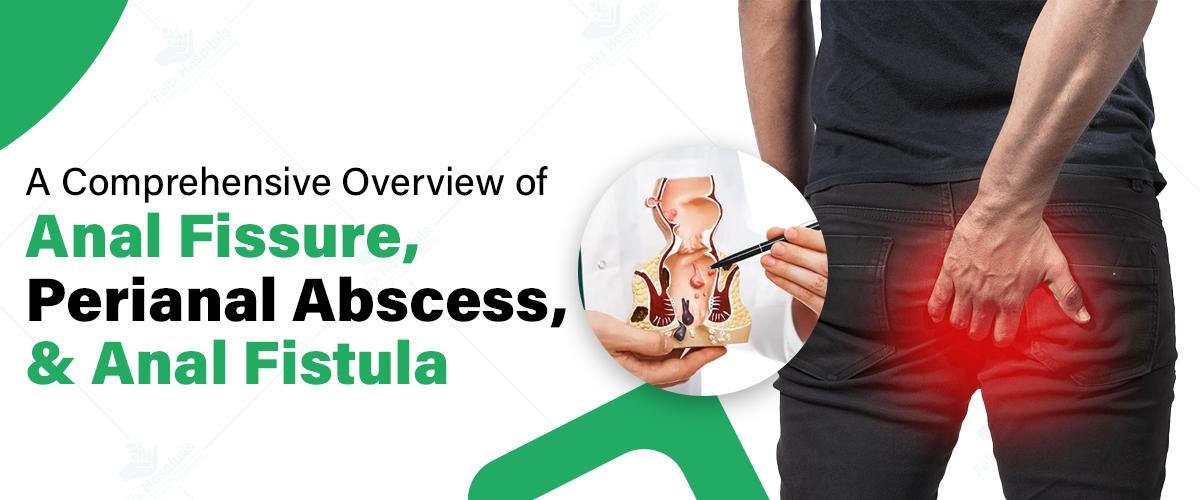
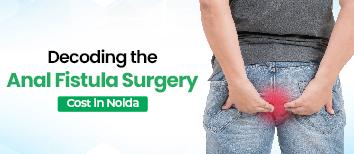
Anorectal disorders affect millions globally, with anal fissures, perianal abscesses, and anal fistulas being particularly impactful. For those experiencing anorectal disorders, timely care is crucial. Understanding these conditions helps individuals recognize symptoms early. By seeking treatment at the best anal fissure hospital or specialized center, patients can receive expert care, improving their overall well-being and quality of life.
Experiencing discomfort from perianal disorders? Reach out to us at +91 9667064100 for effective treatment and care!
Definition and Causes
An anal fissure is a small tear in the lining of the anus. This condition often results from passing hard or large stools, which can stretch and damage the delicate anal tissue. Other factors contributing to Anal Fissures include chronic constipation, diarrhea, childbirth, and inflammatory bowel diseases.
Symptoms
The primary symptom of an anal fissure is sharp pain during bowel movements, which may persist for several hours afterward. Patients may also notice bright red blood on toilet paper or in the toilet bowl. Some individuals experience itching or burning sensations around the anus.
Diagnosis
Healthcare providers typically diagnose anal fissures through a visual examination of the anal area. In some cases, a digital rectal examination may be necessary to rule out other conditions.
Treatment
Conservative management is often the first line of treatment for anal fissures. This includes:
1. Increasing fiber intake and staying hydrated to soften stools
2. Using warm sitz baths to promote healing and relieve pain
3. Applying topical ointments containing nitrates or calcium channel blockers to improve blood flow and promote healing.
In cases where conservative measures fail, botulinum toxin injections or surgical procedures like lateral internal sphincterotomy may be considered.
Definition and Causes
A perianal abscess is a collection of pus near the anus, usually resulting from an infection of the anal glands. These glands can become blocked, leading to the accumulation of bacteria and the formation of an abscess. Risk factors include obesity, diabetes, inflammatory bowel disease, and a compromised immune system.
Symptoms
Patients with perianal abscesses often experience:
1. Severe pain and swelling around the anus
2. Redness and warmth in the affected area
3. Fever and chills
4. Difficulty sitting or having bowel movements
Diagnosis
Diagnosis typically involves a physical examination and, in some cases, imaging studies like CT scans or MRI to determine the extent of the abscess.
Treatment
The primary treatment for perianal abscesses is incision and drainage, a minor surgical procedure performed under local anesthesia. This involves making a small cut to allow the pus to drain. Antibiotics may be prescribed if there are signs of systemic infection or in patients with certain risk factors.
Definition and Causes
An anal fistula is an abnormal tunnel connecting the anal canal to the skin near the anus. It often develops as a complication of a perianal abscess, with about 50% of abscess patients later developing a fistula. Other causes of developing an anal fistula include Crohn's disease, tuberculosis, and rarely, cancer.
Symptoms
Anal fistulas can cause:
1. Persistent drainage of pus or blood from an opening near the anus
2. Pain and swelling in the anal area
3. Irritation of the surrounding skin
4. Difficulty controlling bowel movements in some cases
Diagnosis
Diagnosing an anal fistula typically starts with a comprehensive physical examination and may include further diagnostic tests, such as:
1. Fistulography: An X-ray procedure using contrast material to visualize the fistula.
2. Endoanal Ultrasound: Utilizes high-frequency sound waves to produce detailed images of the anal canal.
3. MRI: Provides detailed imaging to evaluate the extent and trajectory of the fistula.
Treatment
Treatment for anal fistulas usually involves surgery. The specific procedure depends on the location and complexity of the fistula. Common surgical options include:
1. Fistulotomy: Opening the fistula tract to allow it to heal from the inside out.
2. Advancement flap procedure: Using nearby tissue to cover the internal opening of the fistula.
3. LIFT procedure (Ligation of Intersphincteric Fistula Tract): Accessing the fistula through the space between the internal and external anal sphincters.
4. Seton placement: Inserting a thin, flexible cord through the fistula to allow gradual healing.
In some cases, fibrin glue or collagen plugs may be used to seal the fistula tract, although these methods have variable success rates.
While not all anorectal disorders can be prevented, certain lifestyle modifications can reduce the risk of developing these conditions:
1. Maintain a high-fiber diet and stay well-hydrated to promote regular, soft bowel movements
2. Practice good anal hygiene, including gentle cleaning with warm water
3. Avoid straining during bowel movements
4. Exercise regularly to promote overall digestive health
5. Manage underlying conditions like inflammatory bowel disease or diabetes effectively
For individuals with recurring anorectal issues, working closely with a healthcare provider to develop a personalized management plan is crucial.
Dr. Ritesh Agarwal
Dr. Ritesh Agarwal, with 15 years of experience, specializes in treating various anorectal disorders and and his expertise includes open and laparoscopic procedures such as Gallbladder Removal, hernia repair, and non-cancerous breast surgeries, as well as laser treatments for conditions like piles, fissures, fistulas, and varicose veins.
Anal fissures, perianal abscesses, and anal fistulas are common anorectal disorders that can significantly impact an individual's quality of life. Understanding these conditions, their symptoms, and available treatment options, including the anal Fissure Surgery Treatment Cost, is essential for seeking timely medical attention and achieving optimal outcomes. While these topics may be uncomfortable to discuss, open communication with healthcare providers is crucial for effective management and prevention of complications. By staying informed and proactive about anorectal health, individuals can maintain their overall well-being and minimize the impact of these challenging conditions.
Don’t let perianal issues disrupt your life! Call us at +91 9667064100 or click here for specialized care and get back to feeling your best!
Can a perianal fistula heal itself?
In some cases, a perianal fistula might partially heal on its own, but it often requires medical intervention. Without treatment, it may persist or worsen, potentially leading to complications. Professional evaluation and treatment are typically necessary for complete healing.
Can I manage an anal fissure at home?
Yes, mild anal fissures can often be managed at home with measures like increasing fiber intake, staying hydrated, using over-the-counter pain relievers, and taking sitz baths. Persistent or severe fissures should be evaluated by a healthcare provider for proper treatment.
What are the common complications associated with perianal disorders?
Common complications of perianal disorders include recurrent infections, chronic pain, persistent discharge, and the development of new fistulas or abscesses. These issues can significantly impact quality of life and may require ongoing medical management.
How long does recovery from anal fissure surgery typically take?
Recovery from anal fissure surgery generally takes about 4 to 6 weeks. Most people experience significant relief within a few weeks, but full recovery may vary depending on individual healing rates and adherence to post-operative care.
Are there any non-surgical options for treating anal fistulas?
Yes, non-surgical options for treating anal fistulas include medications, such as antibiotics, and procedures like seton placement to facilitate drainage and promote healing. However, surgery is often necessary for complete resolution and long-term success.
Can anal fissures be prevented with diet alone?
While a high-fiber diet is crucial for preventing anal fissures by reducing constipation and straining, it may not be sufficient on its own. Proper hydration and healthy bowel habits are also important for prevention.

फिशर के कारण, लक्षण और उपाय" एक संक्षिप्त मार्गदर्शिका है जो फिशर (गुदा裂) के विषय में जानकारी प्रदान करती है। इसमें फिशर के प्रमुख कारण, जैसे कब्ज, जन्म, या अन्य स्वास्थ्य समस्याएं

एलबेंडाजोल 400 एक प्रभावी एंटीपैरासिटिक दवा है, जिसका उपयोग मुख्य रूप से शरीर में विभिन्न प्रकार के कृमियों (वार्म) को मारने के लिए किया जाता है। यह दवा विशेष रूप से आंतों में पाई जाने वाली कृमियों को खत्म करने में मदद करती है।
एलबेंडाजोल 400 एक एंटीपैरासिटिक (परजीवीरोधी) दवा है, जिसका उपयोग शरीर में मौजूद विभिन्न प्रकार के कृमियों (वार्म) के संक्रमण के इलाज के लिए किया जाता है। यह दवा विशेष रूप से आंतों में पाई जाने वाली कृमियों को मारने और उन्हें शरीर से बाहर निकालने में मदद करती है। एलबेंडाजोल 400 एक महत्वपूर्ण दवा है, जो कृमियों से जुड़े संक्रमणों के इलाज में बहुत प्रभावी होती है। इसका उपयोग डॉक्टर की सलाह के अनुसार ही किया जाना चाहिए, ताकि इससे अधिकतम लाभ प्राप्त किया जा सके और किसी भी संभावित दुष्प्रभाव से बचा जा सके।
सेहत को लेकर कोई भी सवाल है? हमारे डॉक्टर से तुरंत संपर्क करें और सही सलाह पाएं।
कृमि संक्रमण:
यह दवा विभिन्न प्रकार के कृमि संक्रमणों के इलाज में उपयोग की जाती है, जैसे कि राउंडवार्म, हुकवार्म, और टैपवार्म।
हाइडैटिड रोग :
यह एक गंभीर संक्रमण है जो एक परजीवी कीड़ों द्वारा होता है। इस स्थिति में भी एलबेंडाजोल का उपयोग किया जाता है।
फाइलेरियासिस:
फाइलेरियासिस (Filariasis) एक परजीवी संक्रमण है जो फाइलेरिया नामक कृमि के कारण होता है। यह बीमारी मच्छरों के काटने से फैलती है, जो संक्रमित कृमि के लार्वा को शरीर में पहुंचाते हैं।फाइलेरियासिस मुख्यत उष्णकटिबंधीय और उपोष्णकटिबंधीय क्षेत्रों में पाई जाती है और इसका सबसे आम लक्षण लिंफैटिक प्रणाली की सूजन है, जिसे हाथी पांव (एलिफेंटियासिस) भी कहा जाता है।
एलबेंडाजोल 400 का प्रभाव:
एलबेंडाजोल शरीर में मौजूद कृमियों को मारता है और उन्हें मल के माध्यम से शरीर से बाहर निकालने में मदद करता है। यह दवा कृमियों के कोशिका विभाजन को रोकती है, जिससे उनकी वृद्धि रुक जाती है और अंततः वे मर जाते हैं।
एल्बेंडाजोल 400 मिलीग्राम टैबलेट्स उन व्यक्तियों के लिए निर्धारित की जाती हैं, जिन्हें परजीवी कीड़े के संक्रमण का निदान किया गया हो या जिनमें इस प्रकार के संक्रमण का जोखिम हो। निम्नलिखित समूहों के लोगों को एल्बेंडाजोल लेने की सलाह दी जा सकती है:
परजीवी संक्रमण से ग्रसित मरीज:
जिन व्यक्तियों को आंतों, लिवर (Liver), या अन्य अंगों में कीड़े के संक्रमण का निदान हुआ है, उन्हें उनके उपचार के रूप में एल्बेंडाजोल लेने की सलाह दी जाती है।
एंडेमिक क्षेत्रों में यात्रा करने वाले लोग:
जो लोग उन क्षेत्रों में यात्रा कर रहे हैं जहां परजीवी संक्रमण सामान्य हैं, उन्हें संक्रमण के स्रोतों के संपर्क में आने पर एहतियात के तौर पर या लौटने के बाद उपचार के रूप में एल्बेंडाजोल लेने की सलाह दी जा सकती है।
उच्च जोखिम वाले वातावरण में बच्चे:
जिन क्षेत्रों में कीड़े के संक्रमण आम हैं, वहां स्कूल या सामुदायिक एरिया में बच्चों का संपर्क अधिक होता है। ऐसे मामलों में एल्बेंडाजोल को डीवर्मिंग कार्यक्रमों के हिस्से के रूप में बच्चों को दिया जा सकता है।
विशिष्ट चिकित्सीय स्थितियों वाले व्यक्ति:
ऐसे मरीज जिन्हें न्यूरोसिस्टीसर्कोसिस (Neurocysticercosis) या हाइडेटिड रोग जैसी स्थितियां हैं, जिन्हें उपचार की आवश्यकता होती है, उन्हें स्वास्थ्य सेवा प्रदाता की निगरानी में एल्बेंडाजोल लेना चाहिए।
मास ड्रग एडमिनिस्ट्रेशन कार्यक्रमों के प्रतिभागी:
कुछ क्षेत्रों में सामुदायिक स्तर पर परजीवी संक्रमण को नियंत्रित करने और समाप्त करने के लिए एल्बेंडाजोल को मास ड्रग एडमिनिस्ट्रेशन कार्यक्रमों के हिस्से के रूप में वितरित किया जाता है।
यह महत्वपूर्ण है कि एल्बेंडाजोल को केवल तब ही लिया जाए जब यह किसी स्वास्थ्य सेवा पेशेवर द्वारा निर्धारित किया गया हो, क्योंकि अनुचित उपयोग से दुष्प्रभाव हो सकते हैं या उपचार प्रभावी नहीं हो सकता।
एलबेंडाजोल 400 अभी ऑर्डर करने के लिए संपर्क करें: +919667064100।
एल्बेंडाजोल 400 मिलीग्राम टैबलेट्स की खुराक परजीवी संक्रमण के प्रकार, रोगी की उम्र, वजन, और सामान्य स्वास्थ्य पर निर्भर करती है। इसके उपयोग के लिए निम्नलिखित सामान्य दिशानिर्देश दिए गए हैं:
आंतों के कीड़ों के संक्रमण:
अधिकांश आंतों के कीड़ों के संक्रमण के लिए 400 मिलीग्राम की एक खुराक आमतौर पर पर्याप्त होती है। कुछ मामलों में परजीवियों को पूरी तरह से समाप्त करने के लिए दो सप्ताह बाद दूसरी खुराक की आवश्यकता हो सकती है।
हाइडैटिड रोग:
हाइडैटिड रोग (hydatid disease) के लिए सामान्य खुराक 28 दिनों के कोर्स के लिए दिन में दो बार 400 मिलीग्राम है, इसके बाद 14 दिनों का ब्रेक दिया जाता है। संक्रमण की गंभीरता के आधार पर यह चक्र तीन बार तक दोहराया जा सकता है।
न्यूरोसिस्टिसर्कोसिस:
न्यूरोसिस्टिसर्कोसिस के लिए खुराक आमतौर पर 8 से 30 दिनों तक दिन में दो बार 400 मिलीग्राम होती है, जो मरीज की प्रतिक्रिया और चिकित्सक की सिफारिश के आधार पर तय की जाती है। इसे अक्सर कॉर्टिकोस्टेरॉइड्स और एंटीकॉन्वलसेंट्स के साथ दिया जाता है।
लिम्फेटिक फाइलेरियासिस:
लिम्फेटिक फाइलेरियासिस के लिए 400 मिलीग्राम की एक खुराक वार्षिक रूप से दी जाती है, आमतौर पर अन्य एंटिपैरासिटिक दवाओं जैसे इवरमेक्टिन या डायथाइलकार्बामाज़िन (Diethylcarbamazine) के साथ। यह आवश्यक है कि निर्धारित खुराक का पालन ठीक वैसे ही किया जाए जैसा कि स्वास्थ्य देखभाल पेशेवर द्वारा निर्देशित किया गया हो। दवाओं को भोजन के साथ लेना चाहिए ताकि अवशोषण और प्रभावकारिता बढ़ सके।
हालांकि एल्बेंडाजोल 400 मिलीग्राम टैबलेट्स सामान्य होती हैं फिर भी कुछ सावधानियाँ बरती जानी चाहिए:
गर्भावस्था और स्तनपान:
एल्बेंडाजोल को गर्भावस्था के दौरान (during pregnancy) विशेष रूप से पहली तिमाही में लेने की सलाह नहीं दी जाती है, क्योंकि यह भ्रूण के लिए संभावित जोखिम पैदा कर सकता है। प्रजनन आयु की महिलाओं को उपचार के दौरान और उपचार समाप्त होने के एक महीने बाद तक प्रभावी गर्भनिरोधक का उपयोग करना चाहिए। स्तनपान के दौरान एल्बेंडाजोल का उपयोग भी सावधानी से किया जाना चाहिए, क्योंकि यह स्तन के दूध में जा सकता है।
लिवर फंक्शन:
एल्बेंडाजोल का मेटाबोलिज़म लिवर में होता है, इसलिए इसे लिवर की बीमारी वाले मरीजों में सावधानी के साथ उपयोग किया जाना चाहिए। लंबे समय तक उपचार के दौरान लिवर एंजाइम्स की नियमित निगरानी आवश्यक हो सकती है।
बोन मैरो सप्रेशन:
कभी-कभी एल्बेंडाजोल बोन मैरो सप्रेशन का कारण बन सकता है, जिससे श्वेत रक्त कोशिकाओं की संख्या में कमी हो सकती है। लंबी अवधि के उपचार के दौरान नियमित रूप से रक्त परीक्षण किए जाने चाहिए, विशेष रूप से उन मरीजों में जो लंबे समय तक चिकित्सा ले रहे हैं।
एलर्जी प्रतिक्रियाएं:
जिन मरीजों को एल्बेंडाजोल या अन्य बेंज़िमिडाजोल डेरिवेटिव्स से एलर्जी है, उन्हें इस दवा का उपयोग करने से बचना चाहिए। एल्बेंडाजोल 400 मिलीग्राम टैबलेट्स के सामान्य दुष्प्रभावों में सिरदर्द, मतली, पेट में दर्द, चक्कर आना, और अस्थायी बाल झड़ना शामिल हो सकते हैं। गंभीर दुष्प्रभाव दुर्लभ होते हैं लेकिन इनमें लिवर को नुकसान, गंभीर त्वचा प्रतिक्रियाएं और रक्तस्राव संबंधी असामान्यताएं शामिल हो सकती हैं। कोई भी असामान्य लक्षण दिखने पर तुरंत अपने स्वास्थ्य देखभाल प्रदाता से संपर्क करें।
एलबेंडाजोल 400 में सक्रिय तत्व एलबेंडाजोल होता है। जो एक प्रभावी एंटीपैरासिटिक एजेंट है। यह कृमियों के विकास और प्रसार को रोकता है।
यह दवा टैबलेट और सस्पेंशन (तरल) दोनों रूपों में उपलब्ध होती है, जिससे इसे बच्चों और वयस्कों के लिए उपयुक्त बनाया जा सकता है।
एलबेंडाजोल 400 का उपयोग विभिन्न प्रकार के कृमि संक्रमणों जैसे कि राउंडवर्म, हुकवर्म, पिनवर्म, और टैपवर्म के इलाज में किया जाता है। यह दवा आंतों और शरीर के अन्य भागों में मौजूद कृमियों को मारने में मदद करती है।
एलबेंडाजोल कृमियों के कोशिका विभाजन को रोकने में मदद करता है, जिससे उनकी वृद्धि रुक जाती है और अंततः वह मर जाते हैं। मरे हुए कृमियों को शरीर से मल के माध्यम से बाहर निकाल दिया जाता है।
इस दवा की खुराक संक्रमण के प्रकार रोगी की आयु और वजन पर निर्भर करती है। आमतौर पर यह दवा भोजन के साथ या बाद में ली जाती है ताकि इसका अवशोषण बेहतर हो सके।
डॉ. जगतजोत सिंह गिल 11 से अधिक वर्षों के अनुभव के साथ एक प्रतिष्ठित गैस्ट्रोएंटरोलॉजिस्ट हैं, जो हेपेटोलॉजी और इंटरवेंशनल एंडोस्कोपी में विशेषज्ञता रखते हैं। उनकी विशेषज्ञता जटिल परजीवी कृमि संक्रमणों के निदान और उपचार तक विस्तारित है, विशेष रूप से जो गैस्ट्रोइंटेस्टाइनल ट्रैक्ट और लिवर को प्रभावित करते हैं।
डॉ. अमृत योग डटले 8 से अधिक वर्षों के अनुभव के साथ एक सम्मानित गैस्ट्रोएंटरोलॉजिस्ट और क्रिटिकल केयर विशेषज्ञ हैं, विशेष रूप से परजीवी कृमि संक्रमणों के प्रबंधन में। परजीवी संक्रमणों के कारण होने वाली जटिल लिवर और गैस्ट्रोइंटेस्टाइनल स्थितियों के निदान और उपचार में उनके असाधारण कौशल उन्हें अलग करते हैं।
एलबेंडाजोल 400 पर गैस्ट्रोएंटरोलॉजिस्ट की सलाह के लिए आज ही फ़ोन करें - +919667064100.

बदलती दिनचर्या, गलत खान-पान और तनाव के कारण आज गैस्ट्रिक की समस्या आम होती जा रही है। समय पर इलाज और संतुलित जीवनशैली से इस परेशानी से राहत मिल सकती है।

पीलिया एक ऐसी स्थिति है जिसमें शरीर और आंखों का रंग पीला पड़ जाता है। यह मुख्य रूप से लीवर की खराबी या रक्त में बिलीरुबिन के बढ़ने के कारण होता है।
पेट में अल्सर एक आम लेकिन गंभीर समस्या है जो संक्रमण, अधिक एसिड या दवाओं के कारण होती है। इसके लक्षणों में पेट दर्द, जलन और अपच शामिल हैं। फ़ेलिक्स अस्पताल में इसका प्रभावी उपचार और परामर्श उपलब्ध है।
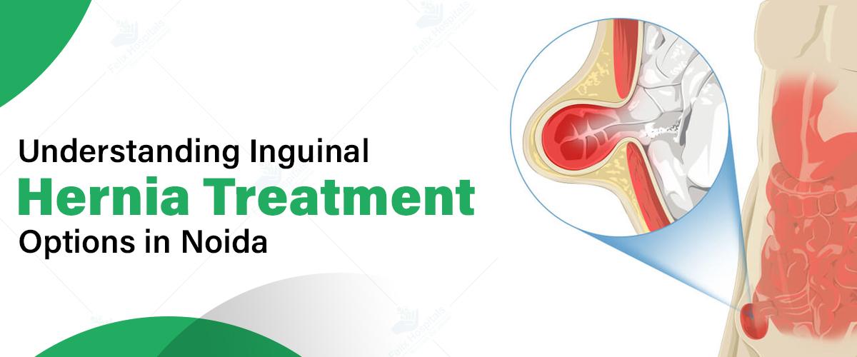
Inguinal hernias are a widespread medical issue impacting millions globally, and Noida, a bustling city in the National Capital Region of India, is no exception. For those affected, understanding the different treatment options available in Noida is essential for making informed healthcare decisions. Exploring these options can lead to better outcomes and a higher quality of life. Additionally, consulting with local specialists can provide personalized advice and support tailored to individual needs.
Struggling with inguinal hernia pain? Call +91 9667064100 or click here for expert treatment and relief!
An inguinal hernia occurs when tissue, such as part of the intestine, protrudes through a weak spot in the abdominal muscles. This type of hernia appears as a bulge in the groin area and can be painful, especially when coughing, bending over, or lifting heavy objects.
The common symptoms for inguinal hernia include:
An inguinal hernia differs from other hernias primarily in its location, causes, symptoms, and treatment:
Location:
Inguinal Hernia | Other Hernias |
| Occurs in the groin area, specifically the inguinal canal. | Can appear in different locations, such as near the belly button (umbilical hernia), through the diaphragm (hiatal hernia), or at a surgical scar (incisional hernia). |
Causes:
Inguinal Hernia | Other Hernias |
| Typically due to increased abdominal pressure and a weakness in the inguinal canal. | Causes vary, like increased abdominal pressure for umbilical hernias or weakening of the diaphragm for hiatal hernias. |
Symptoms:
Inguinal Hernia | Other Hernias |
| Characterized by a bulge in the groin area, with pain or discomfort that worsens with activity. | Symptoms differ; for example, umbilical hernias show as a bulge near the belly button, while hiatal hernias may cause heartburn and difficulty swallowing. |
Treatment:
Inguinal Hernia | Other Hernias |
| Typically requires surgical repair, either open or laparoscopic. | Treatment varies; umbilical hernias may sometimes resolve on their own, while hiatal and incisional hernias often require surgery or other specific management strategies. |
Noida is home to numerous well-equipped hospitals and clinics that are adept at diagnosing inguinal hernias. The diagnostic process generally involves the following steps:
Physical Examination: A healthcare provider will conduct a thorough physical exam, including a visual inspection and palpation of the groin area to identify any noticeable bulges or tenderness.
Imaging Tests: To confirm the diagnosis and assess the hernia's characteristics, imaging tests such as an ultrasound or CT scan may be utilized. These tests help provide a detailed view of the hernia and its impact on surrounding structures.
Felix Hospitals, Noida provides a variety of treatment options for inguinal hernias, catering to different patient needs and preferences. These range from conservative approaches to advanced surgical interventions. Here’s a detailed look at these options:
1. Watchful Waiting
For small, asymptomatic hernias, doctors might recommend a conservative approach known as watchful waiting. This involves:
2. Open Hernia Repair
This traditional surgical technique is commonly available in Noida’s hospitals and involves:
Pros:
Cons:
3. Laparoscopic Hernia Repair
Many hospitals in Noida offer this minimally invasive technique, which includes:
Pros:
Cons:
4. Robotic-Assisted Hernia Repair
Advanced hospitals in Noida are starting to offer robotic-assisted hernia repair, which involves:
Pros:
Cons:
5. Tissue Repair
For certain cases, particularly smaller hernias, some surgeons in Noida may use tissue repair, which involves:
Pros:
Cons:
Each treatment option has its own benefits and limitations, so it is important for patients to discuss these with their healthcare provider to choose the best approach based on their specific condition and circumstances.
Selecting the best treatment for an inguinal hernia involves several key factors. These factors may include:
Size and Location of the Hernia: The hernia’s size and position will determine whether conservative management or a specific surgical approach is needed.
Patient’s Overall Health: The patient’s general health, including any existing medical conditions, influences whether they should opt for surgery or non-surgical methods.
Surgeon’s Expertise: The experience and skill of the surgeon are crucial for successful outcomes, especially with complex cases or advanced surgical techniques.
Available Technology: The technology and facilities at the hospital or clinic in Noida affect the range of treatment options, with more advanced centers offering the latest in minimally invasive and robotic-assisted procedures.
Considering these factors will help in choosing the most effective treatment tailored to individual needs.
When surgery is recommended for an inguinal hernia, patients in Noida should take several important steps to ensure a smooth procedure and recovery:
Undergo Preoperative Tests: Patients will need to complete any pre-operative tests required by their surgeon. These tests may include blood work, imaging studies, and evaluations to assess overall health and ensure there are no underlying issues that could affect the surgery.
Disclose Medications and Supplements: It is crucial to inform the healthcare team about all medications and supplements being taken. This includes prescription drugs, over-the-counter medications, and herbal supplements, as some substances can affect anesthesia or interfere with the surgical procedure.
Follow Fasting Instructions: Adherence to fasting instructions before surgery is essential. Patients will typically be advised not to eat or drink anything for a specific period before the procedure to minimize the risk of complications related to anesthesia and ensure a clearer surgical field.
Arrange for Post-Operative Care and Transportation: Since patients may be groggy or unable to drive after the surgery, it’s important to arrange for someone to transport them home and assist with post-operative care. This includes planning for someone to help with daily activities, medications, and follow-up appointments as needed.
By carefully preparing for these aspects, patients can help ensure a successful surgery and a smoother recovery process.
Dr. Ritesh Agarwal
Dr. Ritesh Agarwal, with 15 years of experience, specializes in both open and laparoscopic hernia surgeries. Practicing at Felix Hospital, Noida, he is adept in various procedures, including laparoscopic hernia repair, gallbladder removal, and laser treatments for proctology issues. Dr. Agarwal is dedicated to providing personalized treatment plans to achieve optimal outcomes for his patients.
Noida provides a wide array of treatment options for inguinal hernias, ranging from conservative management to sophisticated surgical techniques. With its advanced hospitals and skilled surgeons, patients in Noida benefit from top-notch hernia care. By exploring these options and collaborating with healthcare professionals, patients can make well-informed decisions for optimal results. Early diagnosis and timely intervention are crucial for effective hernia management, so if you suspect you have a Hernia, seek prompt evaluation and personalized treatment advice from a healthcare provider in Noida.
Have questions about inguinal hernia treatment? Call +91 9667064100 for expert advice and a customized care plan!
What causes an inguinal hernia?
An inguinal hernia usually results from a combination of muscle weakness and strain. Factors include heavy lifting, chronic coughing, or straining during bowel movements, which push abdominal tissue through a weakened area in the groin.
Is inguinal hernia surgery considered major?
Inguinal hernia surgery is generally considered major due to its invasive nature, though it’s typically performed using minimally invasive techniques. Recovery may involve significant rest and care, but it’s usually well-tolerated with a relatively quick return to normal activities.
Can you resume a normal life after inguinal hernia surgery?
Yes, most people can return to a normal life after inguinal hernia surgery, although a period of rest and gradual reintroduction to physical activities is necessary. Full recovery typically allows for resuming most daily activities without significant limitations.
What are the potential side effects of inguinal hernia surgery?
Side effects of inguinal Hernia Surgery can include pain, swelling, bruising, and infection at the incision site. Some patients might experience numbness, a feeling of heaviness, or recurrence of the hernia. Most side effects are manageable and temporary.
When should surgery be considered for an inguinal hernia?
Surgery should be considered if the hernia causes pain, is growing, or affects daily activities. Urgent surgery may be necessary if there are complications like incarceration or strangulation, where the hernia becomes trapped and its blood supply is compromised.
Can inguinal hernias be prevented?
Preventing inguinal hernias involves avoiding excessive strain on the abdominal muscles, maintaining a healthy weight, and strengthening abdominal muscles through exercise. However, some factors like genetic predisposition may make prevention challenging.
Is it possible for an inguinal hernia to come back after surgery?
Yes, inguinal hernias can recur after surgery, though this is relatively uncommon. Factors such as surgical technique, patient health, and adherence to post-operative care can influence the likelihood of recurrence.
How can I manage pain and discomfort after inguinal hernia surgery?
Pain and discomfort after surgery can be managed with prescribed pain medications, applying ice packs to reduce swelling, and following the doctor’s instructions for activity levels. Gentle movement and rest are important for proper healing.

A hernia typically occurs due to a combination of muscle weakness and strain. This can happen from factors such as aging, injury, heavy lifting, chronic coughing, or conditions like pregnancy and obesity.

Subscribe to our
© Copyright 2026. All Rights Reserved by Felix Healthcare Private Limited
Design and development by :


 +(91)9667064100
 Emergency : +(91)9667064100