 WhatsApp
 WhatsApp


Subscribe to our
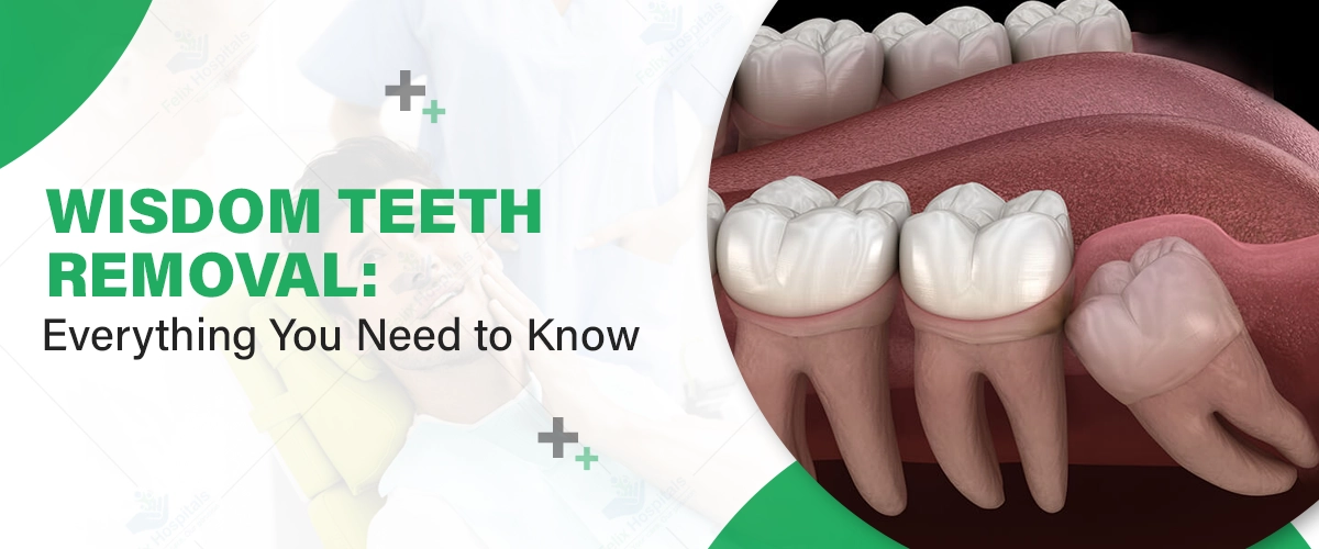
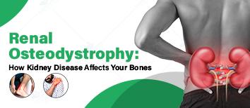
Wisdom teeth, also known as third molars, are the last set to emerge, typically between the ages of 17 and 25. While these teeth served a functional purpose for our ancestors who consumed tough, uncooked foods, they often cause more harm than good in modern humans. Wisdom teeth removal is a common procedure to address oral health issues arising from these teeth.
At the best dental hospital in Noida, patients can expect advanced care and expert guidance when dealing with wisdom teeth. Understanding the signs that necessitate removal and addressing them promptly is essential to prevent complications like infections, crowding, and even damage to surrounding teeth. Timely wisdom tooth removal ensures optimal oral health and reduces the risk of severe conditions.
Book an appointment with our expert dentists and take the first step toward pain-free oral health. Contact us now at +91 9667064100.
Wisdom teeth are the third and final set of molars located in the very back corners of the upper and lower jaws. They are named "wisdom teeth" because they typically appear later in life, during the late teenage years or early adulthood, a time traditionally associated with the gaining of maturity or "wisdom." Most adults have four wisdom teeth—one in each quadrant of the mouth. However, some individuals may have fewer than four, or even none, as wisdom teeth are increasingly considered a vestigial feature of human anatomy.
In early human evolution, wisdom teeth were crucial in chewing raw plants, meats, and roots. Over time, dietary changes and smaller jaw sizes made these teeth redundant. Today, they’re often associated with dental problems rather than benefits.
Wisdom teeth usually emerge between ages 17 and 25. However, in many cases, they fail to erupt properly, leading to a condition known as impaction. Regular dental checkups are vital during this age to monitor their growth and address potential issues.
Impaction
Impacted wisdom teeth grow at an angle or fail to fully emerge from the gums, causing pain, swelling, and potential infections.
Overcrowding
As the last teeth develop, wisdom teeth often lack sufficient space in the jaw, leading to misalignment of surrounding teeth.
Infection or Gum Disease
Partially erupted wisdom teeth can trap food particles and bacteria, resulting in gum inflammation and infection.
Tooth Decay
Difficult-to-reach wisdom teeth are prone to cavities due to inadequate cleaning.
Persistent pain or discomfort in the back of your mouth.
Swelling or redness around the gums.
Difficulty opening your mouth or chewing.
Bad breath or an unpleasant taste caused by trapped food.
If you experience any of these symptoms, consult your dentist promptly to avoid further complications.
A dentist or oral surgeon will assess your wisdom teeth using X-rays to determine their position and identify any issues, such as impaction or misalignment.
Based on the evaluation, your dentist will recommend either monitoring or extracting the teeth. They’ll also discuss the type of anesthesia suitable for your case.
Understanding the steps involved in wisdom teeth removal can help ease any apprehension about the process.
This numbs only the affected area, ensuring you don’t feel pain but remain awake during the procedure. It’s commonly used for straightforward extractions.
In more complex cases, general anesthesia may be administered to put you to sleep throughout the surgery, providing a pain-free experience for more invasive extractions.
Depending on the complexity of your case, the dentist or oral surgeon will administer local or general anesthesia to ensure comfort during the procedure.
If the wisdom tooth is impacted or trapped beneath the gum, an incision is made to expose the tooth for removal.
The tooth is carefully extracted. In some cases, it may be broken into smaller pieces to simplify the removal process and minimize damage to surrounding tissue.
The extraction site is thoroughly cleaned to remove any debris or infection risks, and sutures may be placed to promote healing and protect the area.
This straightforward approach ensures that the procedure is both effective and tailored to the patient’s needs, minimizing discomfort and aiding recovery.
The surgery typically lasts 45 minutes to an hour, depending on the complexity of the case.
First 24 Hours: Swelling and mild bleeding are normal.
1 to 3 Days: Pain peaks but is manageable with prescribed medications.
1 to 2 Weeks: Swelling subsides, and the extraction site begins to heal.
Swelling around the cheeks and jaw.
Bruising or minor bleeding.
Mild fever or fatigue.
Your dentist may prescribe pain relievers, anti-inflammatory drugs, and antibiotics to promote recovery and prevent complications.
Use ice packs on your cheeks to reduce swelling.
Eat soft foods like yogurt, mashed potatoes, and soups.
Rinse your mouth gently with salt water after 24 hours.
Follow your dentist’s post-operative instructions meticulously.
Avoid using straws, as the suction can dislodge blood clots and cause dry sockets.
Refrain from smoking or consuming alcohol.
Skip hard, crunchy, or spicy foods that may irritate the extraction site.
Dry Socket
A painful condition where the blood clot at the extraction site is dislodged. Avoid vigorous rinsing, smoking, or drinking through straws to minimize the risk.
Nerve Damage
Although rare, improper removal may lead to temporary or permanent nerve damage, affecting sensation in the lips, tongue, or jaw.
Signs of Infection
Look out for symptoms such as:
Persistent fever.
Increased redness or swelling around the extraction site.
Pus discharge or foul-smelling breath.
If you notice any of these signs, contact your dentist immediately.
Warning Signs
Persistent pain or discomfort in the back of your mouth.
Difficulty opening your mouth.
Swelling or redness that doesn’t subside.
Bad breath or an unpleasant taste that doesn’t improve.
Routine checkups can help detect wisdom teeth issues early, allowing for timely intervention and preventing complications.
The cost of wisdom teeth removal can vary significantly depending on several factors. Understanding these factors can help you plan for the procedure and make informed decisions.
Whether you need one tooth removed or all four, the number of teeth significantly affects the overall cost. Removing multiple teeth at once may be more cost-effective than scheduling separate extractions.
The choice of anesthesia—local, sedation, or general—plays a major role in determining the cost. While local anesthesia is typically the most affordable, sedation and general anesthesia provide added comfort, albeit at a higher price.
Simple extractions are less expensive, but impacted or partially erupted wisdom teeth often require surgical procedures, which can increase the cost.
Costs may also vary depending on the geographic location of the clinic. Urban areas and premium dental centers may charge higher fees compared to smaller towns or standard clinics.
At our wisdom tooth extraction hospital, you’ll find a team of highly skilled professionals dedicated to ensuring the best outcomes:
Dr. Aditi Narad: Renowned for her expertise in painless extractions and patient-focused care.
Dr. Aditi Sharma: Specializes in managing complex cases of impaction with precision.
Dr. Shinja Dixit: Known for her meticulous approach to oral surgery and compassionate care.
Dr. Migha Singhla: Expert in post-operative management and ensuring quick recovery.
Ready for expert care and a smooth recovery? Click Here to book your appointment online today!
Addressing wisdom teeth issues promptly can prevent severe oral health complications and improve your quality of life. Professional dental care ensures a safe and efficient removal process, minimizing risks and promoting quick recovery.
If you’re experiencing discomfort or suspect you may need wisdom teeth removal, consult a trusted Best dentist in Noida. Regular checkups and timely intervention can help maintain optimal oral health and save you from unnecessary complications.
Q.1. What happens if I ignore impacted wisdom teeth?
Ans: Impacted wisdom teeth can lead to severe complications like infections, cyst formation, jawbone damage, and crowding of other teeth. Prompt evaluation and treatment are critical to avoiding these issues.
Q.2. How can I tell if my wisdom teeth are impacted without X-rays?
Ans: Symptoms like swelling, pain, difficulty opening your mouth, or bad breath can indicate an impaction. However, an X-ray is the most accurate way to diagnose the condition.
Q. 3. Is wisdom teeth removal covered by dental insurance?
Ans: Coverage depends on your insurance provider and plan. Many policies include wisdom teeth extraction under basic dental surgery, especially if medically necessary.
Q. 4. How long does it take to recover from wisdom teeth removal?.
Ans: Most people recover within 1-2 weeks, but healing time can vary based on the complexity of the extraction and adherence to post-operative care instructions.
Q. 5. Can I delay wisdom teeth removal if they are not causing pain?
Ans: Even if you’re not experiencing pain, delayed removal of problematic wisdom teeth can lead to complications later, such as damage to neighboring teeth or an increased risk of infection.
Q. 6. Are there alternatives to extraction for wisdom teeth issues?
Ans: In some cases, dentists may monitor wisdom teeth or prescribe antibiotics for minor infections. However, extraction is often the most effective solution for impacted or problematic teeth.
Q,7. What foods should I avoid after wisdom teeth removal?
Ans: Avoid hard, crunchy, or spicy foods and beverages like soda or coffee during the initial recovery period. Stick to soft foods and follow your dentist’s recommendations.
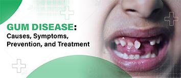
Gum disease, also known as periodontal disease, is an inflammatory condition that affects the tissues surrounding the teeth.
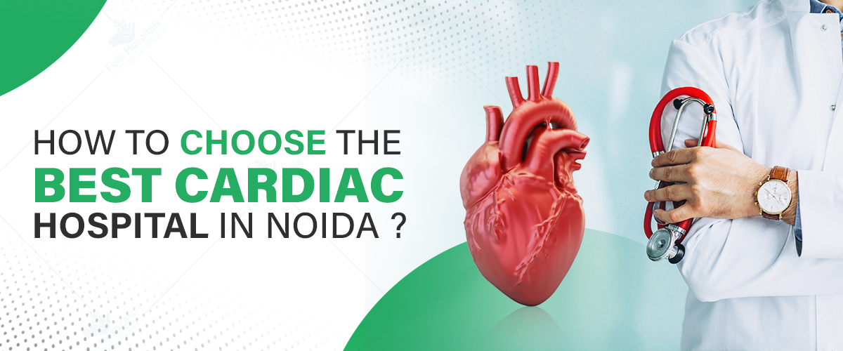
Selecting the right cardiac hospital is crucial for ensuring high-quality treatment and positive health outcomes, especially with the rising prevalence of heart disease. The best cardiac hospital in Noida should offer advanced facilities, experienced specialists, and comprehensive care programs. Key factors to consider include the expertise of cardiologists and surgeons, state-of-the-art diagnostic tools, cutting-edge treatments, and a focus on patient education and rehabilitation. Choosing the right hospital ensures you or your loved one receives the best care for both treatment and long-term heart health.
Have more questions about our cardiac services? Contact Us for More Information by Calling +91 9667064100.
To choose the best cardiology hospital, here are the key factors you should consider:
The expertise of cardiologists plays a significant role in determining the quality of cardiac care. Look for hospitals that have:
Highly qualified and experienced cardiac specialists.
A team of interventional cardiologists, cardiac surgeons, and electrophysiologists who can handle complex procedures.
Positive patient testimonials and success stories reflect the hospital’s track record in treating cardiac patients.
A top-tier cardiac hospital should offer advanced diagnostic and treatment options, including:
ECG, echocardiography, angiography, and other state-of-the-art diagnostic tools.
Cutting-edge treatments such as angioplasty, bypass surgery, and valve replacement.
Minimally invasive procedures that ensure faster recovery and reduced hospital stays.
Cardiac emergencies require prompt medical attention. The hospital should have:
24/7 emergency care and ambulance services for quick response.
A well-equipped Cardiac Intensive Care Unit (ICU) with advanced life-saving equipment.
Efficient protocols for managing heart attacks and other critical conditions.
Preventive and rehabilitative cardiac care is essential for long-term heart health. The hospital should offer:
Preventive cardiology programs to detect risks early and prevent complications.
Cardiac rehabilitation services to aid recovery after surgery or treatment.
Lifestyle management and dietary counseling to help patients maintain a heart-healthy lifestyle.
Comfortable and hygienic hospital infrastructure is essential for a smooth recovery. Consider:
Modern medical facilities and well-maintained hygiene standards.
Comfortable patient rooms with essential support services.
Insurance acceptance and affordable treatment packages for financial ease.
Ensure that the hospital has a good reputation and proper accreditations, such as:
NABH and JCI accreditation, which confirm adherence to global healthcare standards.
Awards and recognitions for excellence in cardiology.
Online reviews and patient ratings that reflect the hospital’s credibility and service quality.
The cost of cardiac treatment varies based on the procedure and hospital. Consider:
Comparative cost analysis of procedures like Angioplasty, bypass surgery, and valve replacement.
Availability of affordable yet high-quality treatment options.
Insurance coverage and cashless treatment facilities for financial convenience.
If you are searching for the Best Cardiologists in Noida, Felix Hospital offers expert cardiac care with renowned specialists, including:
Dr. Rahul Arora – An expert in interventional cardiology and advanced heart procedures.
Dr. Virendra Singh – A leading heart specialist with expertise in preventive and rehabilitative cardiology. Felix Hospital provides world-class cardiac care at competitive costs, making it an excellent choice for heart patients.
Book a consultation with our top cardiologists at Felix Hospital to get personalized care and treatment tailored to your needs.
Choosing the right cardiac hospital involves evaluating multiple factors, including the expertise of doctors, advanced treatment options, emergency care facilities, and cost-effectiveness. Prioritizing your heart health by selecting the Best Cardiac Hospital in Noida can ensure optimal outcomes and a healthier life. If you’re considering heart treatment, consult top specialists at Felix Hospital. The Heart Surgery Cost in Noida varies based on the procedure, but Felix Hospital offers high-quality, affordable care.
1. What are the signs that I need to consult a cardiologist?
Ans: Signs such as chest pain, shortness of breath, dizziness, or a family history of heart disease should prompt a visit to a cardiologist for a thorough evaluation.
2. How do I know if a hospital is equipped to handle complex cardiac surgeries?
Ans: Look for hospitals that offer advanced procedures like bypass surgery, valve replacement, and angioplasty, with experienced cardiologists and specialized surgical teams.
3. What is the difference between interventional cardiology and cardiac surgery?
Ans: Interventional cardiology focuses on minimally invasive procedures like angioplasty to treat heart conditions, while cardiac surgery typically involves open-heart procedures such as bypass or valve replacement.
4. What role does cardiac rehabilitation play in recovery?
Ans: Cardiac rehabilitation is a crucial part of recovery after heart surgery or treatment, helping patients regain strength, improve cardiovascular health, and adopt heart-healthy lifestyle changes.
5. Are minimally invasive heart procedures available in Noida?
Ans: Yes, many leading cardiac hospitals in Noida, including Felix Hospital, offer minimally invasive heart procedures that reduce recovery time and hospital stays.
6. How can I manage heart disease through lifestyle changes?
Ans: Lifestyle management, including regular exercise, a healthy diet, and stress management, plays an important role in controlling heart disease and preventing further complications.
7. What should I consider when evaluating the cost of heart surgery in Noida?
Ans: When evaluating heart surgery costs, consider the procedure type, the hospital’s reputation, the expertise of the doctors, and whether insurance coverage is available for cashless treatment.

In Noida hospitals, recognized for being among the best heart health hospitals in Noida, are at the forefront of offering critical care for heart-related issues.
हार्ट अटैक के मुख्य लक्षण, कारण और तुरंत किए जाने वाले उपचार के बारे में जानें। समय पर पहचान और सही इलाज से जीवन बचाया जा सकता है।
Arrhythmia is a condition where the heart beats irregularly—too fast, too slow, or with an uneven rhythm. It can feel like a fluttering, racing, or skipped heartbeat.

Child surgery plays a crucial role in pediatric healthcare, addressing congenital disorders, injuries, and critical conditions in young patients. With advancements in medical science, surgical outcomes have improved significantly, ensuring safer procedures and faster recovery. If you are looking for the best child surgery hospital in Noida, it is essential to choose a facility equipped with modern technology and experienced pediatric surgeons. Specialized pediatric surgeons understand the unique needs of children, ensuring effective treatment and compassionate care.
Trust Felix Hospital for safe, advanced, and compassionate child surgery treatments. Contact us today at +91 9667064100.
Children’s bodies are still growing and developing, requiring specialized surgical techniques tailored to their unique anatomy. Pediatric surgeons undergo extensive training to address conditions specific to infants and children, making their expertise vital in ensuring the best possible outcomes.
Laparoscopic and robotic-assisted surgeries are widely used in pediatric procedures, offering faster recovery, minimal scarring, and reduced pain. These techniques also contribute to shorter hospital stays, making the recovery process smoother for both children and their families.
Fetal surgery is one of the most advanced aspects of Top Facts about Paediatric and Neonatal Surgery, allowing doctors to operate on babies while they are still in the womb. This cutting-edge approach has successfully treated conditions like spina bifida, congenital heart defects, and other life-threatening abnormalities, giving newborns a healthier start in life.
Due to their rapid cell regeneration, children recover faster from surgeries compared to adults. Proper post-operative care, including a nutritious diet and physical therapy, further enhances healing, ensuring a quicker return to normal activities.
Administering anesthesia to children requires specialized knowledge and techniques. Pediatric anesthesiologists use age-appropriate dosages and modern monitoring systems to minimize risks and ensure a safe surgical experience.
Certain surgical procedures, such as tonsillectomy, hernia repair, appendectomy, and cleft lip surgery, are more frequently performed in children. Early detection and timely treatment lead to better long-term outcomes, reducing complications as they grow.
A multidisciplinary approach is often necessary for complex cases. Pediatric surgeons collaborate with neonatologists, cardiologists, orthopedic specialists, and other medical professionals to provide comprehensive care tailored to each child’s unique needs.
Hospitals prioritize child-friendly environments to ease anxiety before surgery. Techniques like play therapy, storytelling, and counseling help young patients feel more comfortable and prepared for their procedures.
A well-balanced diet accelerates healing and strengthens immunity post-surgery. Pediatric dietitians provide customized nutrition plans to support recovery and promote overall health in young patients.
Emergency situations like appendicitis, trauma, and congenital defects require immediate surgical intervention. Rapid-response pediatric surgical teams ensure timely and effective treatments, improving survival rates and recovery outcomes.
The field of Top Facts about Paediatric and Neonatal Surgery is continuously evolving with advancements in regenerative medicine, artificial intelligence, and robotic-assisted procedures. These innovations are making surgeries safer, more precise, and more effective in treating complex pediatric conditions.
For parents seeking expert pediatric surgical care, Felix Hospital offers a team of highly skilled specialists, including:
If you are searching for the best pediatric surgeon in Noida, Felix Hospital provides advanced surgical treatments with compassionate care to ensure the best outcomes for your child.
Visit our website and schedule an appointment with the best paediatrician in Noida for expert guidance and a personalized treatment plan.
Felix Hospital — Best pediatric surgery in hospital in Noida, delivering specialized pediatric care for newborns, infants, and children with compassion and excellence.
Pediatric surgery has significantly evolved, improving survival rates and recovery times. Selecting an experienced pediatric surgeon ensures the best care and outcomes for young patients. If you are concerned about child surgery treatment cost in Noida, it is advisable to consult a specialized hospital that offers comprehensive and affordable pediatric surgical care. With continuous medical advancements, the future of pediatric surgery looks even more promising, providing safer and more effective treatment options for children worldwide.
Q- What makes pediatric surgery different from adult surgery?
Ans- Pediatric surgeries require specialized techniques as children’s bodies are still developing. Pediatric surgeons undergo extensive training to handle child-specific conditions safely.
Q- Are minimally invasive surgeries safe for children?
Ans- Yes, laparoscopic and robotic-assisted surgeries are widely used in pediatric procedures, ensuring faster recovery, minimal scarring, and reduced pain for young patients.
Q- Can newborns undergo surgery immediately after birth?
Ans- Yes, neonatal surgeries can be performed soon after birth, or even before birth in some cases, to correct life-threatening conditions like congenital heart defects and spina bifida.
Q- How is anesthesia managed in child surgeries?
Ans- Pediatric anesthesiologists use specialized techniques and age-specific dosages to ensure a safe and pain-free surgical experience with minimal complications.
Q- What are the most common pediatric surgeries?
Ans- Some of the most common child surgeries include tonsillectomy, hernia repair, appendectomy, cleft lip correction, and congenital defect repairs.
Q- How can parents prepare their child for surgery?
Ans- Hospitals provide child-friendly environments with play therapy and counseling to help ease anxiety. Parents can also explain the process in a reassuring manner.
Q- How much does child surgery treatment cost in Noida?
Ans- The cost varies based on the type of surgery, hospital facilities, and surgeon expertise. Felix Hospital offers affordable and comprehensive pediatric surgical care.
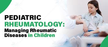
Pediatric rheumatology focuses on diagnosing and treating autoimmune and inflammatory disorders affecting children’s joints, muscles, and connective tissues.

Every parent wants their child to become a successful, confident, and good human being. A child is like a sponge, which can take any shape and can absorb almost everything they see/learn.

10 Reasons why a child may not grow

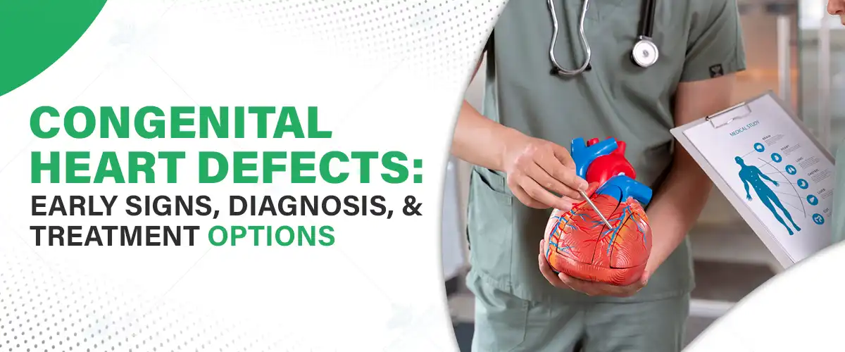
Congenital heart defects (CHDs) are a group of structural abnormalities in the heart that occur during fetal development. These defects can vary significantly in severity and can complicate both physical health and quality of life. Understanding CHD is crucial for early diagnosis and effective treatment. This article aims to shed light on various aspects of congenital heart defects, offering insights into their nature, scientific basis, diagnosis, and treatment. For individuals seeking advanced cardiac care, accessing the Best Heart Hospital in Noida can ensure comprehensive diagnosis, expert treatment, and the latest medical innovations for managing CHDs effectively.
Take the first step towards managing your heart health by visiting our online booking platform. Call us today at +91 9667064100.
Congenital heart defects are present at birth and can result from a variety of factors. They are among the most common types of birth defects, affecting approximately 1 in 100 live births worldwide. CHDs can lead to difficulties in the heart's ability to pump blood, which in turn can impact overall health and wellness.
What are Congenital Heart Defects?
Congenital heart defects encompass a wide range of heart malformations, including abnormal structures, holes between heart chambers, and issues with blood vessels. The term 'congenital' signifies that these defects are intrinsic, emerging during the development of the fetus. Some individuals may experience mild forms of a defect that require little to no treatment, while others may face life-threatening issues necessitating immediate medical intervention.
Types of Congenital Heart Defects
CHDs can be categorized into several types, each with distinct characteristics. Common types include:
Each type has unique consequences on the heart's functionality and health. Families affected by CHD need to understand the specific type impacting their child, as it can influence treatment and management options. For instance, while some defects may resolve on their own as a child grows, others may require surgical intervention or ongoing medical care to ensure proper heart function and overall well-being.
The exact cause of congenital heart defects (CHDs) is often unknown, but several factors may contribute to their development:
Other risk factors include:
Careful monitoring during pregnancy and consultation with healthcare providers can help identify at-risk pregnancies. Additionally, preconception counseling is beneficial for:
Advances in genetic testing and prenatal screening techniques are improving the early detection of CHDs. Research continues to explore how genetic and environmental factors interact, leading to better prevention and intervention strategies.
Congenital heart defects not only pose challenges in diagnosis and treatment but also raise important questions about their underlying scientific mechanisms. Understanding the physiology and genetic influences behind CHD helps in developing better prevention and management strategies.
The Role of Genetics
Genetics plays a significant role in congenital heart defects. Some cases are associated with:
Genetic counseling can help families:
Ongoing research is also exploring:
Congenital heart defects can affect the heart’s structure and function, leading to various complications. The severity of these effects varies:
Regular heart function monitoring is essential for:
Advanced imaging techniques, such as:
These tools allow healthcare providers to tailor treatments for improved patient outcomes.
Individuals with CHD may face additional health challenges, such as:
In complex cases, lifelong care is often required. Multidisciplinary healthcare teams—consisting of pediatricians, cardiologists, and other specialists—play a crucial role in managing these conditions.
Additionally, the psychological impact of living with a heart defect should not be overlooked. Patients and families can benefit from:
These support systems help individuals manage their condition effectively while improving their overall quality of life.
Accurate diagnosis of congenital heart defects is essential for determining the optimal course of action. Advances in medical imaging and technological developments have significantly improved the early detection of CHDs.
Healthcare providers may use several prenatal detection methods, including ultrasound and echocardiography, to identify potential congenital heart defects. These diagnostic tools can reveal structural abnormalities that may indicate a heart defect, allowing for informed decision-making and planning for any necessary interventions prior to or at birth.
After birth, healthcare providers often utilize a combination of clinical assessments and diagnostic imaging techniques to confirm congenital heart defects. These may include chest X-rays, electrocardiograms (ECGs), and further echocardiography to evaluate the heart's function and structure.
Interpreting diagnostic results requires careful assessment by trained medical professionals. The complexity of congenital heart defects necessitates a thorough understanding of each defect's implications for heart function and overall health.
Treatment for congenital heart defects is tailored based on the specific type and severity of the defect, as well as the patient's overall health. Options can range from non-surgical interventions to complex surgical procedures.
In some cases, non-surgical interventions may be sufficient to manage congenital heart defects. These can include medications to improve heart function and manage symptoms, as well as catheter-based therapies that can repair certain defects without the need for open-heart surgery.
For more severe congenital heart defects, surgical intervention may be necessary to repair structural abnormalities and restore optimal heart function. Procedures can range from minimally invasive techniques to complex surgeries, such as open-heart surgery.
Long-term management of congenital heart defects involves ongoing evaluations to monitor heart function, manage associated health issues, and provide psychosocial support. Individuals with CHDs often require a lifetime of follow-up care from specialized healthcare providers.
Educational support and community resources can also play a significant role in enhancing the quality of life for individuals with congenital heart defects. Empowering patients and families through education fosters a better understanding of the condition and encourages proactive management of health and wellness.
Felix Hospital is home to some of the best cardiologists specializing in Congenital Heart Defects (CHDs), ensuring that patients receive expert diagnosis, treatment, and lifelong care. Our team includes:
With state-of-the-art technology, a multidisciplinary approach, and affiliation with the best heart hospital in Noida, Felix Hospital ensures comprehensive care for congenital heart defects, from early diagnosis to long-term management.
Book an Appointment today and experience the highest standard of care at our state-of-the-art facility in Noida.
Congenital heart defects are complex conditions with multi-faceted implications. Awareness, early detection, and tailored treatment strategies are pivotal for ensuring the best possible outcomes for individuals affected by these heart abnormalities. The journey from diagnosis to treatment requires comprehensive support and a collaborative approach to healthcare. Additionally, for those requiring surgical intervention, understanding the Heart Surgery Cost in Noida is essential in planning the best course of treatment. Access to advanced cardiac care and experienced specialists can make a significant difference in improving long-term heart health and overall well-being.
Q- Can congenital heart defects be detected before birth, and how accurate are prenatal screenings?
Ans- Yes, congenital heart defects can often be detected through fetal echocardiography during pregnancy. While accuracy varies depending on the defect, advanced imaging techniques can identify many structural abnormalities before birth, allowing for early planning and intervention.
Q- Are children with mild congenital heart defects at risk for complications later in life?
Ans- Even mild congenital heart defects can lead to long-term complications such as arrhythmias, high blood pressure, or reduced exercise capacity. Regular follow-ups with a cardiologist are essential to monitor any potential changes in heart function.
Q- How does congenital heart disease impact a child's growth and development?
Ans- Some children with CHDs may experience delayed growth and development due to reduced oxygen supply or complications like pulmonary hypertension. Proper medical care, nutrition, and therapy can help manage these challenges.
Q- Is there a link between maternal infections and congenital heart defects?
Ans- Yes, certain maternal infections, such as rubella during pregnancy, have been linked to congenital heart defects. Vaccination and prenatal care can reduce the risk of infections that may contribute to CHDs.
Q- Can lifestyle modifications help manage congenital heart defects without surgery?
Ans- While lifestyle changes alone cannot correct structural defects, they can help manage symptoms and improve overall heart health. Strategies include maintaining a heart-healthy diet, avoiding tobacco and alcohol exposure, and staying physically active within safe limits.
Q- What are the latest advancements in the treatment of congenital heart defects?
Ans- Recent advancements include minimally invasive catheter-based procedures, 3D-printed heart models for surgical planning, and regenerative therapies aimed at repairing damaged heart tissue. These innovations are improving outcomes and reducing recovery times.
Q- How does genetic counseling help families with a history of congenital heart defects?
Ans- Genetic counseling provides families with insights into inherited risks, available genetic tests, and potential preventive measures for future pregnancies. It can help parents make informed decisions about family planning and prenatal care.
A stent procedure can be life-saving for those with blocked arteries. This guide walks you through the stent experience—from the angioplasty process to post-procedure recovery and lifestyle changes.
Find the best cardiologist for your heart care with these essential tips and expert advice.

Millions of individuals across the globe suffer from chronic joint pain, severely limiting activities of daily living. Regardless of the cause of the pain - age, trauma, or another medical condition - joint pain will interfere with your life. Cartilage degeneration is one of the largest perpetrators of joint pain, an ongoing degenerative process resulting in pain and decreased mobility. As we move on to the significance of managing chronic joint pain, it is important to know how newer therapies like cartilage regeneration are promising long-term healing. We at Felix Hospital are orthopedic specialists and use state-of-the-art therapies for our patients to regain their mobility and enhance their lifestyle.
Call Now: +91 9667064100 to learn more and book your consultation!
Chronic joint pain is a nagging ache that lasts for weeks, months, or years. It typically arises as a result of some underlying condition, like osteoarthritis, injury, and autoimmune disease, that leads to inflammation and cartilage wear and tear — the shock-absorbing tissue that cushions our joints. Some of the common reasons for chronic joint pain are:
This ongoing pain can restrict motion, decrease activity, and lead to emotional difficulties like frustration and helplessness.
Cartilage is a smooth, elastic tissue that covers the ends of bones in our joints, allowing smooth, frictionless motion. Its main job is to protect against wear and tear, cushioning and providing shock absorption. Cartilage may be damaged or progressively worn down by injury, overuse, or age. Degeneration is followed by pain, stiffness, and loss of joint motion. When the cartilage is depleted, bones rub against each other, leading to inflammation and increased pain.
Symptoms
Chronic joint pain may manifest itself in various ways depending on what is causing it, but common presentations are:
These may have a detrimental impact on the body and emotional function so minor daily activities become unfeasible.
Causes
Chronic joint pain may be caused by many factors, and determining the cause is critical to cure it effectively. Some of the frequent causes are:
Understanding why people experience chronic joint pain is the secret to knowing what type of relief will be gained from treatment.
Risk Factors
Some risk factors are associated with a greater chance of getting joint pain, such as:
Identification of such risk factors can aid in effective management and prevention of joint pain.
Complications
Joint pain, if left untreated, can lead to various complications that affect physical and mental well-being. These include:
Medical Management (Medical and Non-Surgical Treatment)
Even though joint pain is not always reversible, there are treatments to help manage it. According to the level of pain intensity, other alternatives may be advised:
1. Simple At-Home Remedies:
2. Exercise:
3. Weight Loss:
4. Medication:
5. Topical Treatments:
6. Dietary Supplements:
7. Additional Options:
Surgical Management
If medications do not work, surgery can be performed to end the pain once and for all:
1. Arthroscopy:
2. Joint Fusion:
3. Osteotomy:
4. Joint Replacement:
Cartilage regeneration addresses the root of joint pain—cartilage degeneration—directly. Through stimulating new cartilage tissue growth, regenerative treatments reduce pain, re-establish joint function, and increase mobility. Patients who undergo these therapies tend to experience:
There are several studies and clinical data that prove the efficacy of cartilage repair in offering long-term pain relief to patients with chronic joint pain. Different patients have gained considerable increases in mobility, with others even evading the need for traumatic joint replacement surgery.
While cartilage regeneration treatments are safe, as with any treatment, there are some risks. These include the risk of infection, allergic reaction, or cartilage failure to regenerate. Success in regenerative medicine also varies based on many factors, such as the age of the patient, the severity of damage to the cartilage, and the overall health. Qualified consultation with an expert orthopedic specialist should be conducted to develop the most favorable treatment plan for your condition.
Pricing of the regenerative treatment varies depending on the type of therapy, where the facility is located, and the qualifications of the practitioner. Stem cell and PRP are costlier compared to conventional care but provide sustainable benefits with fewer side effects. In India, cartilage regeneration therapy prices range from ₹50,000 to ₹2,50,000, depending on the treatment chosen. Certain of these therapies may be insured, so it is best to inquire about funding and financing.
Here at Felix Hospital, we boast the best team of orthopedic specialists with experience in addressing chronic joint pain and providing high-level regenerative solutions. Our expertly trained professionals have a personal connection with patients and formulate customized treatment programs that precisely fulfill the needs of every patient.
With their collective experience and novel treatment modalities, Felix Hospital is dedicated to delivering the best possible care for patients in need of relief from chronic joint pain.
Book an appointment with our expert orthopedic specialists at Felix Hospital and explore personalized regenerative treatments that can help restore your joint health.
Cartilage regeneration is a new and promising alternative for patients experiencing chronic joint pain due to cartilage degeneration. Thanks to the developments in regenerative medicine therapies, patients are now able to experience long-lasting pain relief, improved mobility, and improved quality of life. If you have chronic joint pain and would like an individualized treatment plan, speak to the professionals at Felix Hospital to discuss the optimal choice for your condition. Our staff is here to support you every step of the way to long-term relief and enhanced joint health.
1. What is cartilage regeneration, and how does it help with joint pain?
Ans: Cartilage regeneration uses new treatments like stem cell therapy and PRP to activate the body's natural repair and regenerative capacity of the damaged cartilage. This helps reduce joint pain, enhance mobility, and restore joint function.
2. How long does it take to see results from cartilage regeneration treatments?
Ans: The recovery time depends on the extent of cartilage damage and treatment received. In most cases, the patients can expect joint function and pain improvement within weeks to months following the treatment.
3. Is cartilage regeneration suitable for everyone with joint pain?
Ans: While cartilage regeneration is optimistic for the majority, it won't be possible for everyone. Your age, extent of cartilage loss, and general well-being will determine if you would be a good candidate for regenerative treatments.
4. What is the difference between stem cell therapy and PRP therapy for cartilage regeneration?
Ans: Stem cell therapy uses stem cells to regenerate cartilage, while PRP therapy uses concentrated platelets from your own blood to release growth factors that promote tissue healing. Both are effective, but stem cell therapy may offer more long-term results.
5. How do I know if I need cartilage regeneration treatment or if traditional treatments will work?
Ans: A thorough assessment by an orthopedic specialist will determine the best course of treatment. If traditional treatments like medication and physical therapy aren’t providing relief, regenerative therapies may be considered.
6. What are the risks associated with cartilage regeneration treatments?
Ans: While generally safe, there are some risks, such as infection, allergic reactions, or the possibility of the cartilage not regenerating fully. It’s important to discuss these risks with your orthopedic specialist before proceeding with treatment.
7. Does insurance cover cartilage regeneration treatments?
Ans: Insurance coverage for regenerative treatments like stem cell therapy and PRP can vary. It’s best to check with your insurance provider and inquire about coverage options before starting treatment at Felix Hospital.

Bone fractures are common injuries from accidents or falls. While many heal well, complications can arise, affecting recovery. Learn more about these issues and treatments.
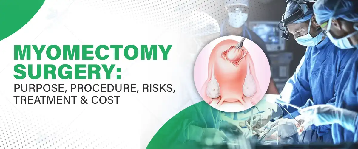
Myomectomy is a specialized surgery that involves the removal of fibroids from the uterus without removing the uterus. It is a significant therapy for those women who experience symptoms like heavy menstrual flow, pelvic pressure, or infertility caused by fibroids. Unlike a hysterectomy, during which the uterus is removed, myomectomy does not interfere with the fertility of women, thus this is the favored choice for those women who want to become pregnant in the future.
At Felix Hospital, we provide expert consultation on complex myomectomy procedures with the finest contemporary facilities and individualized care to achieve the best possible outcome for our patients.
Contact Felix Hospital by calling +91 9667064100 now for a detailed discussion on personalized treatment options tailored for you.
Myomectomy is a surgical procedure to remove fibroids from the uterus. Myomectomy is mainly aimed at the removal of fibroids to correct symptoms without a hysterectomy. It is especially suggested for those women who want to preserve fertility or prevent hysterectomy.
Myomectomy refers to a surgery performed to remove uterine fibroids in a way that preserves the integrity of the uterus. Depending upon the size, number, and position of fibroids, appropriate techniques are applied to gain the most effective outcome possible. A close observation of multiple varieties of myomectomy procedures along with advantages is given below:
1. Abdominal Myomectomy
Abdominal myomectomy, or open myomectomy, is cutting the lower abdomen to remove fibroids. It is usually for patients with:
While it offers good exposure to surgeons and is ideal for complicated cases, it has a longer recovery time compared to minimally invasive methods.
2. Laparoscopic Myomectomy
Laparoscopic myomectomy is a less invasive procedure in which one creates small cuts in the abdomen through which a very small camera and laparoscopic devices are passed. The benefits of this method are:
This method is perfect for patients who have smaller or comparatively smaller-sized fibroids not deeply embedded in the uterine wall.
3. Robotic-Assisted Myomectomy
A myomectomy robot utilizes sophisticated robotic technology to maximize accuracy and control during the operation. The surgery is conducted in a similar way to laparoscopic surgery but with added advantages like:
Robotic myomectomy is an excellent choice for those patients who need soft surgical repair, especially fibroids that are difficult to remove by standard methods.
4. Hysteroscopic Myomectomy
Hysteroscopic myomectomy is a non-surgical method to excise fibroids within the uterine cavity. The procedure is done with a hysteroscope, a thin, flexible tube with a camera, passed through the vagina and cervix. The benefits are:
This is most appropriate for patients with submucosal fibroids (fibroids that develop within the uterine cavity).
The type of myomectomy procedure to use depends on the size, number, and position of fibroids, as well as the overall health of the patient and whether or not future fertility is desired. A consultation with a specialist can assist in determining the most appropriate surgical technique for the best outcome.
Benefits of Myomectomy Surgery
Myomectomy is indicated in women with:
Physicians take into account the size, location, and number of fibroids to decide on the most appropriate course of action.
Myomectomy is a surgical procedure to remove uterine fibroids without removal of the uterus. Effective planning, a carefully thought-out surgical technique, and a planned program of rehabilitation assure a successful operation. This is an exhaustive book on what to do before, during, and after surgery.
Pre-Surgery Preparation
Before myomectomy, patients undergo some steps necessary to make the surgery safe and successful:
Preoperative Instructions: We may ask you to discontinue certain medications, adhere to dietary restrictions, or take medications in a scheduled manner to shrink the fibroid size before surgery.
Anesthesia and Surgical Procedure
The anesthesia used during surgery depends on the myomectomy procedure and the patient's health:
Post-Surgical Recovery
The recovery time will depend on the type of myomectomy that the patient has undergone:
Post-Surgery Care
Myomectomy is safe in most cases, but risks and complications can be:
Low risk at Felix Hospital with expert surgeons and sophisticated technology.
Myomectomy surgery cost is based on:
We provide quality without compromising on prices at Felix Hospital. We also provide insurance coverage and accept flexible payment plans*.
Felix Hospital has a highly professional and experienced team of gynecologists dealing with various specialties of women's health. All our experts are dedicated to delivering the best quality care so that all patients get highly individualized treatment directed toward their specific needs. Our top gynecological team includes:
If you are looking for advanced surgical care, preservation of fertility, or minimally invasive procedures, our specialist gynecologists are there to walk you through each step of the process.
Book a consultation with our expert gynecologists at Felix Hospital today. Let us help you find the best treatment option for your needs.
Myomectomy surgery is a life-altering procedure for women with uterine fibroids, allowing for excellent symptom relief from complaints like heavy menstruation, pelvic pain, and pressure. Other than symptom relief, it is also fertility-saving, making it the best option for women who want to hold on to their reproductive health. With the full information of surgical technology and techniques, such as laparoscopic and robot-assisted myomectomy, patients can now look forward to quicker recovery rates and less likelihood of complications. If you are looking for a myomectomy, then you must approach the specialists at Felix Hospital, where you will be treated with individualized care, the latest facilities, and top-notch medical treatment that suits your requirements.
1. How do I know if I'm a candidate for myomectomy surgery?
Ans: Myomectomy is normally recommended in females who come with signs like heavy menstrual bleeding, pelvic pain, or infertility due to fibroids. A thorough evaluation, like ultrasounds or MRIs, will confirm your suitability for this surgery.
2. Can myomectomy surgery guarantee that my fibroids will never return?
Ans: Although myomectomy successfully eliminates fibroids, there is always a possibility of recurrence in the long term when new fibroids form. Your gynecologist will advise you on the most effective preventive measures and postoperative management.
3. What are the benefits of robotic-assisted myomectomy compared to traditional surgery?
Ans: Robotic-assisted myomectomy provides accuracy and minimally invasive surgery that can result in faster recovery times, less scarring, and fewer complications compared to traditional open surgery.
4. Is myomectomy surgery safe in women who have multiple fibroids?
Ans: Yes, myomectomy can be performed in women who have more than one fibroid, although the type of surgery can be dependent on how and where the fibroids are situated and also how big they are. Your gynecologist will evaluate you and decide the best surgery that will suit your condition.
5. Can I get pregnant after a myomectomy?
Ans: Myomectomy is a procedure that spares fertility, and most women can become pregnant following surgery. Success with pregnancy, however, is contingent on the size of the fibroids, their location, and their general reproductive status.
6. When may I return to normal activities after myomectomy surgery?
Ans: Recovery time will also differ based on the complexity of the surgery. For laparoscopic surgeries, you can return to normal activities within 2-4 weeks, but in more complex cases such as abdominal myomectomy, recovery might take 6 weeks.
7. What can I anticipate when following up after myomectomy surgery?
Ans: You will be monitored in follow-up visits after surgery to check on your healing, address any complications, and take care of any questions about fibroid recurrence or planning a pregnancy. Follow-up visits reassure you that the healing process is going along normally.
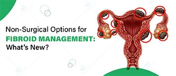
Explore the latest non-surgical treatments for fibroid management. Learn about advanced options to relieve symptoms and improve your quality of life.
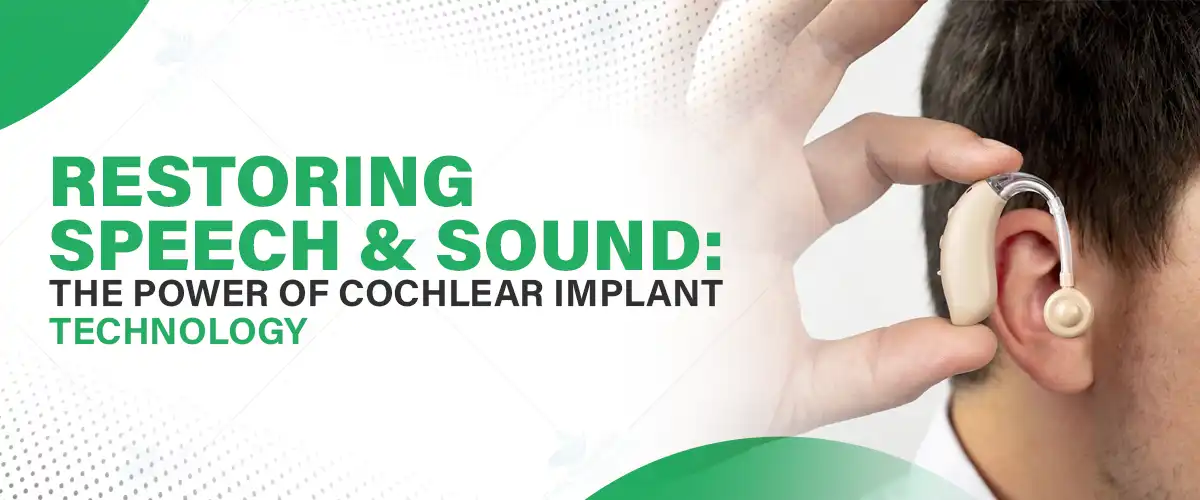
Hearing is an essential sense that connects us to the world, allowing us to communicate, engage in conversations, and enjoy the beauty of sound. For individuals with hearing impairments, the loss of this ability can create significant barriers in daily life. Hearing loss affects millions worldwide, making communication difficult and impacting relationships, employment opportunities, and emotional well-being. Without proper treatment, individuals with profound hearing loss may experience social isolation. Seeking treatment at the best hospital for cochlear implant surgery can open new possibilities for those with severe hearing impairment.
Cochlear implants have emerged as a revolutionary solution for individuals with severe to profound hearing loss. Unlike traditional hearing aids, which amplify sound, cochlear implants work by directly stimulating the auditory nerve, enabling individuals to perceive sound once again. This technology plays a crucial role in restoring speech and sound, offering life-changing benefits.
Schedule a consultation with our expert ENT specialists at Felix Hospital and take the first step towards restoring speech and sound. Call us now at +91 9667064100.
What Are Cochlear Implants?
Cochlear implants are advanced medical devices designed to bypass damaged parts of the inner ear and directly stimulate the auditory nerve. Unlike hearing aids, which only amplify sounds, cochlear implants convert sound into electrical signals, allowing the brain to interpret them as meaningful audio information.
How Do Cochlear Implants Work?
The cochlear implant system consists of two main components:
This process enables individuals to experience sound, facilitating the restoration of speech and sound for those with profound hearing impairment.
Candidates for Cochlear Implants
Cochlear implants are recommended for individuals who:
Age Considerations
Cochlear implants are effective for both children and adults.
Medical Evaluation Process
Candidates must undergo a thorough medical evaluation, including:
Pre-Surgery Preparation
Patients considering cochlear implants undergo detailed consultations to set realistic expectations and understand potential risks and benefits.
The Surgical Procedure
Cochlear implant surgery is a safe and minimally invasive procedure. The key steps include:
Activation and Rehabilitation
Post-surgery, the implant is activated after healing, a moment often described as life-changing. Rehabilitation includes:
Restoring Speech and Sound
Cochlear implants significantly improve speech recognition and the ability to hear various sounds, from conversations to environmental noises.
Improving Quality of Life
Users report enhanced confidence, social interactions, and a renewed sense of independence, reducing feelings of isolation and depression.
Success Stories
Many individuals have transformed their lives through cochlear implants. From young children developing speech to adults re-entering the workforce, these devices make a significant impact in restoring speech and sound.
Innovations in Design and Functionality
Recent advancements have made cochlear implants:
Future Trends
Ongoing research aims to:
Adjusting to the Implant
Adapting to cochlear implants requires time, patience, and auditory training.
Cost and Accessibility
Maintenance and Follow-Up
Regular check-ups ensure optimal device performance. Users may require:
Felix Hospital is committed to providing world-class care for individuals with hearing loss, offering advanced cochlear implant surgery to restore hearing and improve quality of life. Whether you're in Noida Sector 137 or Greater Noida Gamma 1, our expert ENT specialists ensure precise treatment and a smooth recovery journey.
Our Expert ENT Specialists Include:
Dr. Kunwar Parwez – A leading ENT surgeon specializing in cochlear implants, known for his expertise in advanced hearing restoration techniques and patient-centered care.
Dr. Arvinder Pal Singh – An expert in ENT microsurgeries and implantable hearing devices, delivering customized treatment plans for patients with severe hearing loss.
Dr. Arjun Saini – Highly skilled in cochlear implantation and audiological rehabilitation, ensuring successful outcomes and improved auditory function.
With our dedicated specialists and state-of-the-art technology, Felix Hospital offers the best care for cochlear implant surgery, helping patients regain the joy of hearing.
Experience the Power of Cochlear Implants! Don’t let hearing loss hold you back. Book your consultation today!
Cochlear implants offer a revolutionary way to restore speech and sound, transforming lives by enabling clear communication and a richer auditory experience. If you or a loved one struggles with severe hearing loss, consult professionals to explore this life-changing option. The cochlear implant surgery cost in Noida is a worthwhile investment in regaining the ability to hear and connect with the world.
With continuous technological advancements, cochlear implants will become even more accessible and effective, offering hope to millions affected by hearing loss worldwide.
Q1- How soon after surgery will my cochlear implant be activated?
ANS: The implant is typically activated 2–4 weeks after surgery to allow proper healing. During the activation, an audiologist will program the device and guide you through initial sound adjustments.
Q2- Will I regain normal hearing with a cochlear implant?
ANS: While cochlear implants don’t fully restore natural hearing, they significantly improve speech recognition and sound perception, helping individuals communicate effectively and recognize environmental sounds.
Q3- Can both ears be implanted at the same time?
ANS: Yes, bilateral cochlear implants can be performed for better sound localization and improved speech understanding, especially in noisy environments. However, eligibility depends on medical evaluations.
Q4- Is cochlear implant surgery painful?
ANS: The procedure is performed under general anesthesia, so you won’t feel pain during surgery. Post-surgery discomfort is mild and manageable with prescribed medication, and most patients recover within a few days.
Q5- Will I need speech therapy after getting a cochlear implant?
ANS: Yes, speech therapy and auditory training are crucial for adapting to the new way of hearing. This helps the brain process sounds effectively and improves speech clarity over time.
Q6- What is the success rate of cochlear implants?
ANS: The success rate is very high, especially for individuals who receive the implant early. Most users experience significant improvements in hearing ability, communication skills, and overall quality of life.
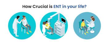
ENT health is essential for proper breathing, hearing, speech, and balance. Early detection and expert care ensure a better quality of life and prevent chronic complications.
Congenital Heart Defects (CHDs) are structural abnormalities of the heart present at birth. They can range from simple conditions that require minimal intervention to complex defects that need immediate medical attention. Early detection and congenital heart failure treatment play a vital role in improving patient outcomes. Raising awareness about CHD helps in timely diagnosis and appropriate medical intervention, ensuring a better quality of life for affected individuals.
Felix Hospital offers state-of-the-art medical facilities and specialized care for CHD patients. Call us Now at +91 9667064100.
CHDs refer to a group of heart conditions that develop before birth, affecting the heart’s structure and function. These defects can involve the heart’s walls, valves, or blood vessels. Some common types include:
Prevalence: CHDs are the most common birth defect, affecting nearly 1 in 100 newborns worldwide.
Recognizing the symptoms of CHDs in infants, children, and undiagnosed adults can lead to timely treatment and improved outcomes.
In Infants and Children:
In Adults (If Undiagnosed in Childhood):
Since CHDs vary in severity, early recognition of these symptoms allows for prompt medical evaluation and intervention.
CHDs develop due to genetic and environmental factors affecting fetal heart formation.
Genetic Factors:
Environmental Factors:
Other Risk Factors:
Proper prenatal care and a healthy lifestyle can help reduce the risk of CHDs.
Prenatal Screening:
Postnatal Diagnostic Tests:
If a CHD is suspected after birth, doctors may use:
Specialist Consultation:
A pediatric cardiologist is essential for an accurate diagnosis and treatment plan.
The treatment approach depends on the type and severity of the heart defect.
Medical Management:
Surgical Interventions:
Minimally Invasive Procedures:
Long-Term Care:
Timely diagnosis and treatment significantly improve survival rates and enhance the quality of life for individuals with CHDs. Advances in pediatric cardiology have made it possible for children with heart defects to live long, active lives with proper care.
At Felix Hospital, expert pediatric cardiologists provide cutting-edge diagnostic tools and personalized treatment plans, ensuring the best outcomes for patients. Routine monitoring is essential for lifelong heart health.
Individuals with CHDs can lead fulfilling lives with the right care and support.
For Children:
For Adults:
Emotional Well-being:
Many CHD patients experience stress or anxiety related to their condition. Support groups and counseling can help both patients and families navigate these challenges.
For expert care in congenital heart failure treatment, consult with:
Felix Hospital provides state-of-the-art facilities and comprehensive care for CHD patients.
If you notice any symptoms of CHD in yourself or your child, Book your appointment and consult with our expert for advanced treatment options.
Early detection, accurate diagnosis, and advanced congenital heart failure treatment are key to managing congenital heart defects effectively. If you or your loved one is experiencing any heart-related symptoms, consult the best cardiologists at Felix Hospital.
For more information on congenital heart failure treatment costs in Noida, contact Felix Hospital today. Raising awareness and seeking timely medical help can make a life-changing difference.
Q- Can congenital heart defects be detected before birth?
Ans- Yes, CHDs can be detected through fetal echocardiography during pregnancy, usually between 18-24 weeks of gestation. Early detection helps in planning treatment strategies after birth.
Q- Are all congenital heart defects life-threatening?
Ans- Not all CHDs are severe. Some minor defects, like small atrial septal defects (ASD), may close on their own, while complex conditions like hypoplastic left heart syndrome (HLHS) require immediate intervention.
Q- Is surgery the only treatment option for congenital heart defects?
Ans- No, treatment depends on the severity of the defect. Some mild CHDs can be managed with medications and regular monitoring, while others may require catheter-based interventions or surgery.
Q- Can adults develop complications from an undiagnosed congenital heart defect?
Ans- Yes, adults with undiagnosed CHDs may experience arrhythmias, heart failure, stroke, or high blood pressure in the lungs (pulmonary hypertension), requiring late-stage interventions.
Q- How do lifestyle changes help in managing congenital heart defects?
Ans- A heart-healthy diet, regular low-impact exercise, medication adherence, and avoiding smoking and alcohol can help individuals with CHDs maintain heart function and overall well-being.
Q- What is the survival rate for children with congenital heart defects?
Ans- Thanks to advancements in medical technology, over 85% of children with CHDs survive into adulthood with proper medical care, including surgery and lifelong monitoring.
Q- Does congenital heart failure treatment cost in Noida vary by hospital?
Ans- Yes, the cost depends on the complexity of the defect, treatment approach (medications, surgery, or catheter-based procedures), and hospital facilities. Felix Hospital provides affordable, high-quality care for CHD patients.
In Noida hospitals, recognized for being among the best heart health hospitals in Noida, are at the forefront of offering critical care for heart-related issues.
Angioplasty is a common heart procedure used to open blocked arteries and restore proper blood flow.
A stent procedure can be life-saving for those with blocked arteries. This guide walks you through the stent experience—from the angioplasty process to post-procedure recovery and lifestyle changes.

Heart disease remains one of the leading causes of death worldwide, with millions of individuals affected each year. The prevalence of coronary artery disease (CAD), heart attacks, and other heart conditions is a growing concern. Early detection and intervention are key in managing heart disease, which is where diagnostic tools like the CT Angiogram (CTA) come into play. Identifying potential problems before symptoms appear allows for timely treatment, improving the chances of avoiding more severe complications.
When looking for the Best CT Angiogram Hospital in Noida, it's crucial to seek hospital who are well-versed in interpreting complex results and offering personalized care. The CT Angiogram is a highly effective diagnostic procedure used to assess the health of your heart and blood vessels, giving your doctor the critical information needed to make informed decisions about your heart health.
Understanding your Heart Score can help you stay ahead of heart disease. Consult with our expert radiologists by Calling +91 9667064100.
A CT Angiogram (CTA) is a sophisticated imaging test that uses X-rays and a contrast dye to create detailed pictures of your blood vessels. This non-invasive procedure allows doctors to observe the blood flow in the coronary arteries, which supply oxygen-rich blood to the heart. The contrast dye injected during the test highlights any blockages or narrowing of these arteries, making it easier for doctors to identify potential risks of heart disease.
CT Angiograms are specifically used to detect coronary artery disease (CAD), which is one of the most common causes of heart attacks. The ability to visualize the arteries helps identify blockages or other abnormalities that may lead to severe health issues.
Heart disease encompasses a wide range of conditions, with Coronary Artery Disease (CAD) being one of the most prevalent. CAD occurs when the blood vessels that supply the heart become narrowed or blocked, often due to the buildup of plaque. This can lead to chest pain, heart attacks, and other serious complications.
Other heart conditions include heart failure, arrhythmias (irregular heartbeats), and heart valve problems. The risk factors for heart disease include:
Having a heart score can help assess your overall risk of developing heart disease, allowing you to make necessary lifestyle changes and seek appropriate treatment before it's too late.
The CT Angiogram plays a vital role in the early detection of heart disease, particularly in identifying blockages or narrowing of coronary arteries. This test is invaluable because it can detect potential problems even before symptoms appear, which is crucial for effective intervention.
Compared to traditional angiograms, CTA is less invasive and involves less radiation, making it a safer option. It provides clearer images, and the procedure is typically faster, offering an efficient and accurate way to assess the condition of your heart.
By identifying early signs of coronary artery disease, a CT Angiogram can help doctors take preventive measures to avoid heart attacks or strokes, ensuring that patients receive timely treatment.
Your Heart Score is a measure of your risk for coronary artery disease, calculated using the results of a CT Angiogram. This score takes into account the degree of blockage in your coronary arteries and gives you an idea of how likely you are to experience heart-related issues in the future. The Heart Score helps doctors determine the severity of the condition and develop a treatment plan that is tailored to your specific needs.
The Heart Score ranges from low to high risk, based on how much of your coronary arteries are blocked. The higher the score, the greater your risk for heart disease, and the more aggressive your treatment plan may need to be.
The Heart Score is categorized into three main risk levels:
Your doctor will use the Heart Score to guide treatment and preventive measures, helping you maintain a healthy heart.
The primary advantage of a CT Angiogram is the early detection of coronary artery blockages, which can lead to heart attacks or strokes. By catching these issues early, doctors can provide you with a personalized treatment plan that could prevent the need for invasive surgeries later on.
CTA is also non-invasive, so you can avoid more complicated procedures like traditional angiography. With its ability to quickly identify heart disease risks, the CT Angiogram provides patients with peace of mind, knowing they are taking steps toward proactive heart health management.
Several groups of individuals should consider undergoing a CT Angiogram:
If you fall into any of these categories, it's essential to consult with a doctor to assess whether a CT Angiogram is the right diagnostic tool for you.
While the CT Angiogram is generally safe, there are a few risks to consider:
Always consult with your doctor to determine if a CT Angiogram is the best option for your specific health needs.
Before the procedure, you may need to fast for a few hours. You may also be asked to adjust certain medications. During the test, a contrast dye will be injected into your bloodstream, and a CT scan will capture images of your arteries. The test typically lasts 30 to 60 minutes, and you can resume normal activities shortly after.
After the test, your doctor will review the results and discuss any findings with you. If your Heart Score indicates a risk of coronary artery disease, your doctor may recommend further tests or treatments, such as lifestyle changes, medications, or even surgery.
Regular follow-up appointments are essential to monitor your heart health and ensure that you're taking the appropriate steps to prevent heart disease.
Felix Hospital is home to highly skilled radiologists who specialize in CT Angiography, ensuring precise diagnosis and expert care. Our team includes:
With state-of-the-art technology and a patient-centric approach, our radiologists at Felix Hospital provide the highest standard of care for CT Angiography, helping patients take proactive steps toward better heart health.
Don't wait for symptoms to appear—early detection is key to preventing serious heart conditions. Book an appointment online now!
CT Angiograms are crucial in diagnosing and preventing heart disease, helping doctors catch potential problems early and create personalized treatment plans. If you're at risk or experiencing symptoms, it’s important to consult with the CT Angiogram Doctors in Noida to discuss whether this procedure is right for you.
Additionally, understanding your Heart Score can provide valuable insights into your heart health and guide decisions about lifestyle changes, medications, or treatments. Make sure to prioritize regular check-ups, as early detection is key to maintaining a healthy heart. If you're concerned about the CT Angiogram test cost, it's important to discuss this with your healthcare provider, as costs can vary based on the hospital and your specific medical needs.
Q- How does a CT Angiogram compare to a traditional angiogram?
Ans- A CT Angiogram (CTA) is non-invasive and uses X-rays with contrast dye to visualize arteries, whereas a traditional angiogram requires inserting a catheter into an artery. CTA is quicker, less risky, and doesn’t require recovery time like a catheter-based procedure.
Q- Can a CT Angiogram detect heart blockages that haven’t caused symptoms yet?
Ans- Yes, a CT Angiogram can detect early-stage blockages in coronary arteries, even before they cause symptoms like chest pain or shortness of breath. This allows for preventive measures before severe complications arise.
Q- Is a CT Angiogram suitable for individuals with existing heart conditions?
Ans- Yes, CTA is often used for monitoring patients with heart disease, assessing stents, bypass grafts, and identifying new blockages. However, the decision depends on the severity of the condition and your doctor's recommendation.
Q- How accurate is a CT Angiogram in diagnosing coronary artery disease (CAD)?
Ans- A CT Angiogram has over 90% accuracy in detecting coronary artery blockages, making it a highly reliable diagnostic tool for identifying mild to severe CAD.
Q- What precautions should diabetics or kidney patients take before a CT Angiogram?
Ans- Since the contrast dye used in a CT Angiogram can affect kidney function, diabetic and kidney patients should inform their doctor beforehand. Pre-procedure hydration or alternative imaging methods may be recommended.
Q- How often should someone at risk of heart disease undergo a CT Angiogram?
Ans- The frequency depends on individual risk factors such as age, family history, high cholesterol, or previous heart conditions. In general, high-risk patients may need periodic scans every 1-3 years for proactive monitoring.
Q- Can a CT Angiogram replace a treadmill stress test or an ECG?
Ans- A CT Angiogram provides detailed images of arteries, whereas stress tests and ECGs measure heart function and electrical activity. While CTA is more precise in detecting blockages, a combination of tests may be required for a full heart assessment.
Angioplasty is a common heart procedure used to open blocked arteries and restore proper blood flow.
A stent procedure can be life-saving for those with blocked arteries. This guide walks you through the stent experience—from the angioplasty process to post-procedure recovery and lifestyle changes.

Tonsils, small glands located at the back of the throat, play a critical role in the body’s immune system by fighting infections. However, when tonsils become frequently inflamed or enlarged, they can cause chronic health issues. In such cases, removal of the tonsils becomes necessary. The Coblation technique has emerged as a revolutionary method in ENT (Ear, Nose, and Throat) care, providing a safer and more comfortable alternative for tonsil removal.
If you’re searching for the best hospital for tonsil treatment, modern advancements like Coblation ensure effective care with minimal discomfort.
Experience world-class care with Coblation techniques at Felix Hospital. Contact us now at +91 9667064100 to schedule your consultation!
Tonsils are part of the immune system and act as the first line of defense against bacteria and viruses entering through the mouth or nose. Despite their protective role, they can become a source of chronic infections and other complications.
Common reasons for tonsil removal:
Chronic Tonsillitis: Frequent infections causing severe throat pain and fever.
Sleep Apnea: Enlarged tonsils can obstruct the airway, leading to disrupted sleep patterns.
Difficulty Swallowing: Enlarged tonsils may interfere with swallowing or speaking.
Signs your tonsils might need removal:
Persistent sore throat.
Swelling that interferes with breathing.
Recurrent infections unresponsive to antibiotics.
Traditional tonsillectomy methods include cold steel removal, laser treatments, and electrocautery. While effective, these methods often cause significant post-surgical pain, prolonged recovery, and potential complications.
Coblation Technique:
This modern method uses radiofrequency energy to dissolve tonsil tissue with minimal heat, significantly reducing discomfort and recovery time. Unlike traditional techniques, Coblation causes less damage to surrounding tissues, making it an advanced choice for tonsil removal.
Advantages of Coblation:
Reduced pain and discomfort.
Faster healing process.
Minimal bleeding.
Precise removal with less tissue damage.
Coblation offers several benefits that make it a preferred choice for tonsillectomy:
Less Pain: The low-temperature procedure minimizes nerve and tissue trauma.
Faster Recovery: Patients typically resume normal activities sooner than with traditional methods.
Reduced Bleeding: Coblation’s precise approach minimizes blood loss during surgery.
Shorter Procedure Time: The technique is efficient, often requiring less time in the operating room.
The Coblation tonsillectomy is a straightforward and patient-friendly procedure:
Anesthesia: General or local anesthesia is administered to ensure comfort.
Tonsil Removal: A specialized Coblation wand dissolves the tonsil tissue without generating excess heat.
Completion: The procedure usually lasts 30-45 minutes. Most patients can return home the same day.
Recovery from a Coblation tonsillectomy is typically smoother than traditional methods. Here’s what to expect:
Pain Management: Mild painkillers may be prescribed for post-surgery discomfort.
Diet: Stick to soft foods and stay hydrated to aid healing.
Rest: Ample rest is essential for a quick recovery.
Follow-up Care: Regular check-ups ensure optimal healing.
While Coblation is considered safe, patients should be aware of potential risks, including infection or mild bleeding. These risks are significantly reduced when the procedure is performed by an experienced ENT specialist.
Choosing a skilled surgeon at the best hospital for tonsil treatment ensures a safe and successful surgery.
Coblation tonsillectomy is suitable for:
Children and adults experience frequent tonsil infections.
Individuals with sleep apnea or difficulty swallowing due to enlarged tonsils.
Those seeking a minimally invasive and low-pain alternative to traditional surgery.
Patients with certain medical conditions may require additional evaluation before undergoing the procedure.
Our ENT specialists are dedicated to offering world-class care for patients needing tonsil surgery. Whether you’re in Noida Sector 137 or Greater Noida Gamma 1, Felix Hospital provides the Best doctor for tonsils treatment, ensuring expert care and a smooth recovery journey.
Our Expert ENT Specialists Include:
Dr. Kunwar Parwez: A renowned ENT surgeon with extensive experience in Coblation tonsillectomy, known for his precision and compassionate care.
Dr. Arvinder Pal Singh: An expert in advanced ENT procedures, offering tailored treatment plans and exceptional surgical outcomes.
Dr. Arjun Saini: Highly skilled in modern ENT techniques, including Coblation, ensuring minimal discomfort and faster patient recovery.
With a team of dedicated professionals, Felix Hospital ensures you receive the highest standard of care throughout your tonsil removal journey.
Visit Felix Hospital in Noida Sector 137 or Greater Noida Gamma 1 for the best tonsil treatment. Schedule a Consultation for your better care!
The Coblation technique is a modern, effective, and patient-friendly solution for removing tonsils. With reduced pain, faster recovery, and minimal complications, it stands out as a preferred choice for tonsil surgery. If you’re considering tonsillectomy, understanding the tonsil surgery cost and consulting a skilled ENT specialist is the first step to better health.
For expert care, advanced technology, and a patient-centric approach, Felix Hospital is here to guide you. Book your consultation today and take the first step toward a healthier, more comfortable life.
Q1- What makes the Coblation technique better than traditional tonsil removal methods?
ANS: The Coblation technique uses low-temperature radiofrequency energy to dissolve tonsil tissue, resulting in less pain, reduced bleeding, and faster recovery compared to traditional methods like cold steel or laser tonsillectomy.
Q2- How long does it take to recover from Coblation tonsil surgery?
ANS: Most patients recover within 7-10 days, significantly faster than traditional tonsillectomy methods. Following a soft diet and prescribed medications can further ease recovery.
Q3- Are there any dietary restrictions after Coblation tonsil removal?
ANS: Yes, patients are advised to stick to soft, non-spicy foods and stay hydrated during the initial recovery phase. Cold or lukewarm items, such as yogurt or soups, can help soothe the throat.
Q4- Is Coblation tonsillectomy safe for children?
ANS: Absolutely! Coblation is considered a safe and effective method for tonsil removal in children, as it minimizes tissue damage and speeds up recovery, making it less traumatic for young patients.
Q5- How much does tonsil surgery cost at Felix Hospital?
ANS: The cost of tonsil surgery varies based on individual needs and medical conditions. Felix Hospital provides competitive pricing and personalized treatment plans. Contact us for a detailed estimate tailored to your case.
Q6- Can I return to work or school after the Coblation tonsillectomy?
ANS: Most patients can resume their routine activities within a week, though strenuous activities should be avoided for at least two weeks. Follow-up care with your ENT specialist ensures a smooth transition back to your daily life.
ENT health is essential for proper breathing, hearing, speech, and balance. Early detection and expert care ensure a better quality of life and prevent chronic complications.

Subscribe to our
© Copyright 2026. All Rights Reserved by Felix Healthcare Private Limited
Design and development by :


 +(91)9667064100
 Emergency : +(91)9667064100