 WhatsApp
 WhatsApp


Subscribe to our
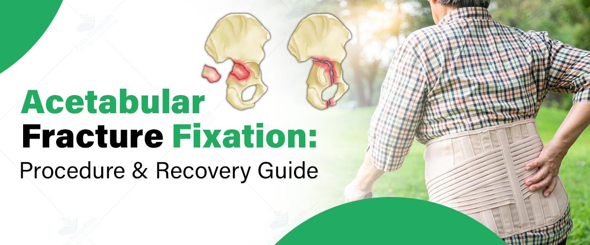
Acetabular fractures involve damage to the socket portion of the hip joint (the acetabulum), which plays a crucial role in maintaining the stability of the hip. This type of fracture is often the result of high-impact trauma, such as car accidents or falls from significant heights. Given the complexity of these fractures, proper fixation is vital for long-term recovery and to prevent complications that could affect mobility and quality of life. Seeking treatment at the best orthopedic hospital in Noida can significantly improve outcomes, as specialized care, advanced imaging, and experienced surgeons are crucial in managing such injuries. Understanding the procedure for acetabular fracture fixation and the recovery timeline is essential for patients to achieve the best possible results and regain their mobility.
Start Your Recovery Journey Now! Contact us at +91 9667064100 with our specialists in Noida and Greater Noida.
An acetabular fracture refers to a break in the acetabulum, which is the cup-like structure in the pelvis that forms the socket of the hip joint. These fractures are typically caused by significant trauma, such as motor vehicle accidents, falls, or sports-related injuries. The acetabulum works in conjunction with the femoral head (the ball portion of the hip joint) to provide a stable and functional hip joint.
There are several types of acetabular fractures, including:
Certain factors increase the risk of acetabular fractures, including:
Surgical fixation is typically necessary when the fracture is displaced or the hip joint is unstable. In cases where the fracture fragments misalign and cannot heal properly on their own, surgery is crucial. Several factors influence the decision to perform surgery, including:
Early intervention plays a key role in the recovery process. Fixing the fracture promptly improves the chances of a full recovery, reduces the risk of complications, and enhances long-term mobility.
Before surgery, the patient undergoes several diagnostic tests, including X-rays, CT scans, and physical exams. These tests help the surgeon assess the severity of the fracture and plan the best approach for fixation.
There are two primary types of fixation methods:
Surgical anesthesia options include general anesthesia, where the patient is fully asleep, or regional anesthesia, where only the lower body is numbed.
Immediately after surgery, the patient is monitored in a recovery room for signs of complications. Pain management is essential in this phase, and the medical team administers medication to keep discomfort under control. The hospital stay typically lasts several days, depending on the complexity of the surgery and the patient’s overall health.
Physical therapy begins as soon as possible to improve joint mobility, muscle strength, and overall functionality. The rehabilitation process is crucial to restoring the hip's full range of motion and preventing long-term complications. Patients are also monitored for potential complications, such as infections, blood clots, or issues with the fixation hardware (e.g., screws or plates).
The recovery timeline for acetabular fracture fixation varies based on the severity of the fracture, the patient’s age, and other health factors. It generally unfolds in three stages:
1. Immediate Post-Surgery Phase (First Few Days to Weeks): During this phase, patients are encouraged to rest and follow instructions regarding weight-bearing restrictions. Crutches or a walker may be required to avoid placing weight on the hip joint. Swelling, bruising, and discomfort are common, and patients typically remain in the hospital for a few days.
2. Short-Term Recovery (6 Weeks to 3 Months): At this stage, physical therapy plays an important role in regaining mobility. Patients gradually begin to increase weight-bearing on the affected hip, with the goal of achieving some level of mobility. X-rays are often performed to ensure the fracture is healing correctly. This phase may involve limited weight-bearing and assisted walking.
3. Long-Term Recovery (6 Months to 1 Year): Complete recovery from an acetabular fracture can take up to one year. During this time, patients work on strengthening the muscles surrounding the hip and improving flexibility. Full weight-bearing activities and a return to normal activity levels can occur, depending on the success of the surgery and the patient’s adherence to rehabilitation protocols.
Physical therapy is essential throughout the recovery process. Early exercises focus on improving flexibility and preventing joint stiffness, while later exercises emphasize building strength in the hip and leg muscles. The rehabilitation team will tailor exercises to the patient's progress and limitations. Full restoration of mobility is typically achieved within 6 to 12 months, but each patient’s timeline may vary.
During recovery, lifestyle adjustments may be necessary, such as avoiding high-impact activities (e.g., running, jumping) that could stress the healing joint. Nutritional considerations are also crucial; calcium and vitamin D-rich foods can promote bone healing. Maintaining bone health through weight-bearing exercises (when appropriate) and fall prevention strategies is key in avoiding future fractures.
Felix Hospitals is proud to offer expert care for acetabular fracture fixation, ensuring the best possible outcomes for patients. Whether you're seeking treatment in Noida or Greater Noida, our team of skilled orthopedic surgeons is equipped to provide personalized care tailored to your needs.
At Felix Hospital in Sector 137, Noida, patients can trust the expertise of our renowned orthopedic specialists:
These professionals bring extensive experience and advanced techniques to ensure a smooth recovery and excellent results.
For those in Greater Noida, Dr. Varun Aggrawal serves patients at Felix Hospital Gamma-1, Greater Noida. Known for his commitment to excellence, Dr. Aggrawal provides world-class orthopedic care to address complex acetabular fractures effectively.
Book an Appointment Today with the best orthopedic surgeons at Felix Hospitals today for expert care and personalized treatment.
Acetabular fracture fixation is a complex procedure, but when performed at a skilled orthopedic center, it offers patients a chance at a full recovery. A well-structured recovery plan that includes surgical intervention, post-operative care, and rehabilitation is crucial for regaining mobility and function. Adhering to medical advice and taking proactive steps during recovery can help patients return to their normal lives and reduce the risk of future complications. Seeking care at the best hospital for Acetabular fracture fixation in Noida ensures that patients receive the expertise and support they need for optimal healing.
1. What is an acetabular fracture?
Ans: An acetabular fracture is a break in the cup-like structure of the pelvis that forms the socket of the hip joint. This type of fracture typically results from high-impact trauma such as car accidents or falls.
2. When is surgery required for an acetabular fracture?
Ans: Surgical fixation is necessary when the fracture is displaced, the hip joint is unstable, or the bones cannot heal properly on their own. Surgery helps restore alignment and ensures proper healing.
3. What are the risks of acetabular fracture surgery?
Ans: While rare, potential risks include infection, blood clots, nerve or blood vessel damage, and issues with the fixation hardware. Choosing an experienced orthopedic surgeon can minimize these risks.
4. How long does it take to recover from acetabular fracture fixation surgery?
Ans: Recovery can take 6 to 12 months, depending on the severity of the fracture, the patient’s age, and adherence to rehabilitation protocols. Physical therapy is crucial for regaining strength and mobility.
5. What should I avoid during recovery from acetabular fracture fixation?
Ans: Avoid high-impact activities, such as running or jumping, until fully healed. Follow your doctor’s instructions regarding weight-bearing restrictions and maintain a diet rich in calcium and vitamin D for bone healing.
6. Can an acetabular fracture heal without surgery?
Ans: In cases where the fracture is non-displaced and the hip joint remains stable, non-surgical treatment may be an option. However, most acetabular fractures require surgical intervention for optimal healing.
7. Why should I choose Felix Hospitals for acetabular fracture fixation?
Ans: Felix Hospitals offers advanced imaging, skilled orthopedic surgeons, and personalized care to ensure the best outcomes for acetabular fractures.

Bone fractures are common injuries from accidents or falls. While many heal well, complications can arise, affecting recovery. Learn more about these issues and treatments.
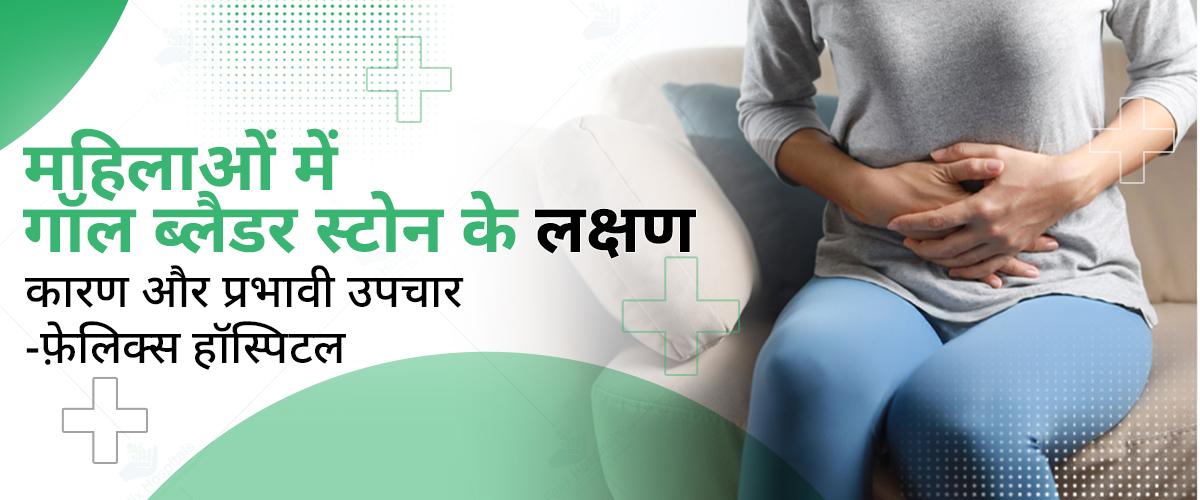
गॉल ब्लैडर (पित्ताशय) में पथरी या स्टोन होना महिलाओं में एक सामान्य समस्या बनती जा रही है। गॉल ब्लैडर, जो पित्त (बाइल) को संग्रहित और नियंत्रित करता है, पाचन में सहायक होता है। लेकिन कई बार यहां कोलेस्ट्रॉल या बिलीरुबिन के क्रिस्टल जमने लगते हैं, जिससे स्टोन बनने की संभावना बढ़ जाती है इसलिए समय रहते आप अच्छे ब्लैडर स्टोन ट्रीटमेंट हॉस्पिटल से समपर्क करें। इस ब्लॉग में हम गॉल ब्लैडर स्टोन के लक्षण, कारण और प्रभावी उपचार के बारे में जानेंगे, ताकि महिलाएं इस समस्या को समय रहते पहचान सकें और इलाज करा सकें।
ज्यादा जानकारी के लिए हमें कॉल करें +91 9667064100.
गॉल ब्लैडर स्टोन (पित्ताशय की पथरी) महिलाओं में एक सामान्य समस्या है, जिसमें गॉल ब्लैडर में पत्थर जैसी संरचनाएं बनने लगती हैं। गॉल ब्लैडर यकृत के नीचे स्थित एक छोटा अंग है, जो पित्त को संग्रहित करता है। यह पित्त वसा के पाचन में मदद करता है। जब बाइल में कोलेस्ट्रॉल, बिलीरुबिन, या अन्य पदार्थों की अधिकता हो जाती है, तो ये पदार्थ ठोस हो कर छोटे पत्थर जैसे रूप में बदल जाते हैं, जिन्हें पित्ताशय की पथरी कहते हैं। गॉल ब्लैडर स्टोन का समय पर उपचार करवाना बहुत जरूरी होता है, अन्यथा यह संक्रमण और गंभीर स्वास्थ्य समस्याओं का कारण बन सकता है।
महिलाओं में गॉल ब्लैडर स्टोन के लक्षण कई प्रकार के हो सकते हैं और अक्सर दर्द या अन्य असुविधाजनक स्थितियों के रूप में सामने आते हैं। यहां कुछ सामान्य लक्षण दिए गए हैं:
यह सबसे आम लक्षण है, जिसे बाइलिक कोलिक कहते हैं। दर्द अचानक आता है और हल्के से लेकर तीव्र तक हो सकता है, जो कुछ मिनटों से लेकर कई घंटों तक रह सकता है।
यह दर्द अक्सर दाएं कंधे और पीठ के दाईं ओर फैल सकता है।
कुछ महिलाओं को भोजन के बाद विशेष रूप से वसा युक्त भोजन के बाद मतली और उल्टी की समस्या हो सकती है।
पथरी से प्रभावित महिलाओं को वसा युक्त भोजन के बाद पेट में भारीपन, सूजन, और गैस की समस्या हो सकती है।
यदि पथरी के कारण संक्रमण हो गया है, तो तेज बुखार, पसीना, और ठंड लगने की समस्या भी हो सकती है।
यदि पथरी बाइल डक्ट (पित्त नलिका) को अवरुद्ध कर देती है, तो त्वचा और आंखों का रंग पीला हो सकता है।
अवरुद्ध पित्त नलिका के कारण, मल का रंग हल्का और यूरिन का रंग गहरा हो सकता है।
गॉल ब्लैडर स्टोन के लक्षणों का अनुभव हर महिला में अलग हो सकता है और गंभीरता में भिन्न हो सकते हैं। अगर लक्षण लगातार और तीव्र हो रहे हों, तो तुरंत चिकित्सा परामर्श लेना जरूरी होता है, क्योंकि इससे संक्रमण या अन्य जटिलताएं उत्पन्न हो सकती हैं।
महिलाओं में गॉल ब्लैडर स्टोन मुख्य रूप से दो प्रकार के होते हैं, जो पत्थर की संरचना और उनकी उत्पत्ति के आधार पर अलग-अलग होते हैं:
ये गॉल ब्लैडर स्टोन का सबसे सामान्य प्रकार हैं, और ज्यादातर महिलाओं में पाए जाते हैं। कोलेस्ट्रॉल स्टोन पीले या हरे रंग के होते हैं। ये तब बनते हैं जब बाइल में कोलेस्ट्रॉल की अधिकता होती है, जो घुल नहीं पाता और ठोस कणों में बदल जाता है। इसका कारण खराब आहार, मोटापा और हार्मोनल बदलाव हो सकता है, जैसे गर्भावस्था के दौरान।
ये गहरे भूरे या काले रंग के होते हैं और तब बनते हैं जब बाइल में बिलीरुबिन की मात्रा बढ़ जाती है। बिलीरुबिन एक पिगमेंट है जो शरीर में लाल रक्त कोशिकाओं के टूटने से बनता है। पिगमेंट स्टोन अक्सर उन महिलाओं में बनते हैं जो लिवर की बीमारियों जैसे सिरोसिस, रक्त विकार जैसे सिकल सेल एनीमिया, या बाइल डक्ट में संक्रमण से पीड़ित होती हैं।
महिलाओं में गॉल ब्लैडर स्टोन के कई कारण हो सकते हैं, जिनमें हार्मोनल बदलाव, खान-पान की आदतें, और जीवनशैली से जुड़े कई कारक शामिल हैं। यहां कुछ प्रमुख कारण दिए गए हैं:
इन कारणों को ध्यान में रखकर यदि महिलाएं अपने खान-पान और जीवनशैली में बदलाव करती हैं, तो गॉल ब्लैडर स्टोन की संभावना को कम किया जा सकता है।
गॉल ब्लैडर स्टोन से बचाव और निदान के लिए कुछ एहतियात और जांच प्रक्रियाएं अपनाई जा सकती हैं। समय पर पहचान और सही उपचार से इस समस्या को रोका और नियंत्रित किया जा सकता है।
वसा और कोलेस्ट्रॉल की मात्रा कम रखें। अधिक तले हुए, वसायुक्त और जंक फूड से बचें। फाइबर युक्त खाद्य पदार्थ जैसे सब्जियां, फल और साबुत अनाज का सेवन करें। चीनी और रिफाइंड कार्बोहाइड्रेट का सेवन सीमित करें।
अधिक वजन गॉल ब्लैडर स्टोन का खतरा बढ़ा सकता है। स्वस्थ तरीके से वजन कम करें और नियमित व्यायाम करें।
नियमित शारीरिक गतिविधि बाइल का प्रवाह बनाए रखती है, जिससे पथरी बनने का खतरा कम होता है।
अनियमित भोजन और अधिक लंबे समय तक भूखे रहना गॉल ब्लैडर स्टोन की संभावना बढ़ा सकता है। सही समय पर भोजन करें।
ये आदतें यकृत और गॉल ब्लैडर की कार्यक्षमता को प्रभावित कर सकती हैं और पथरी की संभावना को बढ़ा सकती हैं।
अगर आप हार्मोनल दवाएं ले रही हैं या गर्भनिरोधक गोलियों का उपयोग कर रही हैं, तो डॉक्टर से परामर्श लें, क्योंकि ये पथरी के खतरे को बढ़ा सकती हैं।
यदि लक्षण महसूस होते हैं, तो डॉक्टर कुछ जांचों की सलाह दे सकते हैं ताकि गॉल ब्लैडर स्टोन का पता लगाया जा सके:
यह सबसे सामान्य और प्रभावी जांच है जो गॉल ब्लैडर में स्टोन की उपस्थिति की पुष्टि करती है। अल्ट्रासाउंड से पथरी का आकार, संख्या, और स्थान ज्ञात होता है।
यह जांच गॉल ब्लैडर की कार्यक्षमता को मापती है और यह पता लगाने में मदद करती है कि बाइल ब्लैडर से सही तरीके से निकल रहा है या नहीं।
सीटी स्कैन से गॉल ब्लैडर और बाइल डक्ट की विस्तार से तस्वीरें ली जाती हैं। इससे छोटे स्टोन भी दिखाई दे सकते हैं।
यह एमआरआई की तरह एक विशेष स्कैन है, जो बाइल डक्ट्स और गॉल ब्लैडर की विस्तृत छवि दिखाता है और जटिल मामलों में उपयोगी होता है।
ब्लड टेस्ट:
ब्लड टेस्ट से संक्रमण, पीलिया, और बाइल डक्ट के अवरोध का पता लगाया जा सकता है। इसमें बिलीरुबिन, लिवर एंजाइम, और सफेद रक्त कोशिकाओं की मात्रा देखी जाती है।
अगर गॉल ब्लैडर स्टोन की पुष्टि हो जाती है और लक्षण गंभीर हैं, तो डॉक्टर सर्जरी की सलाह दे सकते हैं। हल्के लक्षणों वाले मामलों में, लाइफस्टाइल में सुधार और आहार पर ध्यान देकर पथरी को बढ़ने से रोका जा सकता है।
गॉल ब्लैडर स्टोन का जल्दी निदान और उचित बचाव पथरी के कारण होने वाले दर्द और अन्य जटिलताओं को रोक सकता है।
महिलाओं में गॉल ब्लैडर स्टोन का उपचार लक्षणों की गंभीरता, पथरी के आकार, और स्थिति पर निर्भर करता है। यहां कुछ प्रभावी उपचार विकल्प दिए गए हैं:
लेप्रोस्कोपिक कोलेसिस्टेक्टॉमी: यह एक न्यूनतम इनवेसिव सर्जरी है जिसमें पेट में छोटे-छोटे चीरे लगाए जाते हैं और गॉल ब्लैडर को निकाल दिया जाता है। यह सबसे आम और सुरक्षित प्रक्रिया है, जिसमें अस्पताल में कुछ ही दिन रुकना पड़ता है।
यदि लेप्रोस्कोपिक सर्जरी संभव नहीं होती या जटिलताएं होती हैं, तो ओपन सर्जरी का सहारा लिया जाता है। इसमें पेट में बड़ा चीरा लगाया जाता है, जिससे मरीज को ठीक होने में थोड़ा अधिक समय लगता है।
यदि सर्जरी संभव नहीं है, तो कुछ दवाएं (जैसे उर्सोडिऑल या केनोडिऑल) दी जाती हैं जो कोलेस्ट्रॉल स्टोन को घोलने में मदद कर सकती हैं। हालांकि, इस उपचार में समय लगता है और यह छोटे कोलेस्ट्रॉल स्टोन के लिए ही प्रभावी होता है। यह तरीका पिगमेंट स्टोन के लिए उतना कारगर नहीं होता।
अगर पथरी बाइल डक्ट में अटक जाती है और संक्रमण या जटिलताओं का कारण बनती है, तो ईआरसीपी की मदद से इसे हटाया जाता है। इस प्रक्रिया में एंडोस्कोप के माध्यम से बाइल डक्ट से पथरी को बाहर निकाला जाता है। यह एक सर्जरी नहीं है, लेकिन इसमें विशेषज्ञता की आवश्यकता होती है।
यह एक गैर-सर्जिकल प्रक्रिया है जिसमें उच्च-ऊर्जा वाली शॉक वेव्स का उपयोग करके गॉल ब्लैडर स्टोन को छोटे-छोटे टुकड़ों में तोड़ा जाता है, ताकि वे बाइल के साथ निकल सकें। यह प्रक्रिया बहुत आम नहीं है और इसे केवल छोटे कोलेस्ट्रॉल स्टोन के लिए ही उपयोग किया जाता है।
अगर पथरी छोटी है और गंभीर लक्षण नहीं है, तो आहार और जीवनशैली में सुधार से इसे नियंत्रित किया जा सकता है। वसा युक्त खाद्य पदार्थों से बचें, संतुलित आहार लें, और नियमित व्यायाम करें। इससे गॉल ब्लैडर के स्वास्थ्य में सुधार हो सकता है और पथरी के बढ़ने की संभावना कम होती है।
कुछ लोग सेब का रस, सेब का सिरका, या हल्दी का उपयोग करते हैं, जो बाइल फ्लो में सुधार ला सकते हैं। हालांकि, इनका वैज्ञानिक प्रमाण सीमित है, इसलिए किसी भी घरेलू उपचार को अपनाने से पहले डॉक्टर से परामर्श लेना चाहिए।
अगर गॉल ब्लैडर स्टोन का आकार बढ़ रहा है या लक्षण बिगड़ रहे हैं, तो नियमित जांच और डॉक्टर की सलाह पर सर्जरी या अन्य प्रक्रियाएं करवाना सही रहता है।
गॉल ब्लैडर स्टोन का प्रभावी उपचार का चयन स्थिति, लक्षण, और स्वास्थ्य पर निर्भर करता है। सही उपचार व नोएडा में इलाज की कीमत के बारे में जानने के लिए डॉक्टर से परामर्श लेना सबसे महत्वपूर्ण है, ताकि पथरी से होने वाली संभावित जटिलताओं से बचा जा सके।
गॉल ब्लैडर स्टोन के इलाज के लिए कुछ विशेष प्रकार के डॉक्टरों और विशेषज्ञों की आवश्यकता होती है, जो इस स्थिति की पहचान, निदान, और उपचार में मदद करते हैं।
डॉ. रितेश अग्रवाल: फेलिक्स हॉस्पिटल में लैप्रोस्कोपिक सर्जरी में विशेषज्ञता रखने वाले प्रमुख सर्जन हैं, जो मिनिमली इनवेसिव प्रक्रियाओं के जरिए जटिल सर्जिकल मामलों का इलाज करते हैं।
डॉक्टर की सलाह के लिए आज ही फोन करें +91 9667064100.
महिलाओं में गॉल ब्लैडर स्टोन की समस्या आम होती जा रही है, लेकिन इसे पहचानने और सही उपचार के जरिए इससे बचा जा सकता है। समय पर डॉक्टर से परामर्श लेकर सही उपचार करवाना बेहद जरूरी है, ताकि पथरी से होने वाली किसी भी जटिलता को रोका जा सके। स्वस्थ जीवनशैली और नियमित स्वास्थ्य जांच से आप इस समस्या से काफी हद तक बच सकती हैं। इस जानकारी से आपको गॉल ब्लैडर स्टोन के बारे में सही निर्णय लेने में मदद मिलेगी और इस समस्या से निपटने में सहायता मिलेगी।
प्रश्न 1: महिलाओं में गॉल ब्लैडर स्टोन क्यों होते हैं ?
उत्तर: हार्मोनल बदलाव, विशेषकर एस्ट्रोजन का बढ़ा हुआ स्तर, अनुवांशिकता, मोटापा, अधिक कोलेस्ट्रॉल वाला आहार, और तेजी से वजन घटाना महिलाओं में गॉल ब्लैडर स्टोन के सामान्य कारण हैं।
प्रश्न 2: गर्भावस्था में गॉल ब्लैडर स्टोन का खतरा बढ़ता है?
उत्तरः हां, गर्भावस्था में एस्ट्रोजन और प्रोजेस्टेरोन के स्तर में बदलाव होने से गॉल ब्लैडर में कोलेस्ट्रॉल की मात्रा बढ़ सकती है, जिससे स्टोन बनने की संभावना बढ़ जाती है।
प्रश्न 3: क्या गॉल ब्लैडर स्टोन का इलाज बिना सर्जरी के संभव है ?
उत्तर: छोटे कोलेस्ट्रॉल स्टोन के लिए दवाइयां और शॉक वेव लिथोट्रिप्सी जैसी विधियों से इलाज संभव है, लेकिन बड़े स्टोन और गंभीर लक्षणों के लिए सर्जरी की आवश्यकता हो सकती है।
प्रश्न 4: गॉल ब्लैडर स्टोन से कैसे बचा जा सकता है ?
उत्तर: वसा और कोलेस्ट्रॉल युक्त खाद्य पदार्थों का सेवन कम करें, संतुलित आहार लें, नियमित व्यायाम करें, और वजन को नियंत्रित रखें। यह सब गॉल ब्लैडर स्टोन से बचाव में सहायक हो सकता है।
प्रश्न 5: क्या गॉल ब्लैडर हटाने से कोई दुष्प्रभाव होता है ?
उत्तर: गॉल ब्लैडर हटाने के बाद शरीर में बाइल को सीधे छोटी आंत में छोड़ा जाता है, जिससे कुछ लोगों को पाचन में हल्की समस्याएं हो सकती हैं, लेकिन अधिकतर लोग सामान्य जीवन जी सकते हैं।
प्रश्न 6: गॉल ब्लैडर स्टोन की पहचान कैसे होती है ?
उत्तर: अल्ट्रासाउंड, सीटी स्कैन, या एमआरआई जैसी इमेजिंग तकनीक से गॉल ब्लैडर स्टोन की पहचान की जा सकती है। गंभीर मामलों में ईआरसीपी भी की जाती है।
प्रश्न 7: क्या गॉल ब्लैडर स्टोन कैंसर का कारण बन सकता है ?
उत्तर: लंबे समय तक गॉल ब्लैडर में स्टोन रहने से गॉल ब्लैडर कैंसर का जोखिम बढ़ सकता है, लेकिन ऐसा बहुत दुर्लभ होता है। गॉल ब्लैडर स्टोन की सही समय पर पहचान और इलाज से इस खतरे को कम किया जा सकता है।
प्रश्न 8: क्या गॉल ब्लैडर स्टोन के साथ सामान्य जीवन संभव है ?
उत्तर: यदि स्टोन से कोई गंभीर लक्षण नहीं हैं, तो कुछ लोग गॉल ब्लैडर स्टोन के साथ भी सामान्य जीवन जी सकते हैं। लेकिन समय-समय पर डॉक्टर से जांच कराते रहना आवश्यक है ताकि जटिलताओं से बचा जा सके।
Gallbladder stone surgery, or cholecystectomy, is typically performed when gallstones lead to significant health issues.

When faced with a health issue, knowing where to seek care—whether at a polyclinic or a hospital—can be crucial for timely and effective treatment. The choice often depends on factors such as affordability, convenience, type of illness, and access to specialized medical knowledge. Let’s explore the differences between polyclinics and hospitals, helping you understand which option may be best suited for your health needs.
Trust Felix Hospitals for the best care, specialized medical services, and compassionate support during your critical moments by Clicking Here.
A polyclinic is a healthcare facility that offers a range of outpatient services, usually from general practitioners and specialists. It is typically smaller than a hospital but provides comprehensive care, including diagnostic tests, preventive care, and minor procedures. Polyclinics are usually situated in communities or near residential areas, making them accessible for routine consultations and minor health issues.
Services Provided: Routine check-ups, vaccinations, minor surgeries, lab tests, and general consultations.
Doctors and Specialists: Often includes general practitioners, dermatologists, dentists, and other specialists.
Affordability: Polyclinics tend to be more affordable than hospitals, making them a preferred choice for non-urgent care.
Hospitals are large medical facilities equipped to handle a wide range of health conditions, from routine check-ups to emergency care and complex surgeries. Hospitals often include multiple departments with advanced diagnostic and treatment facilities, inpatient care, and specialized intensive care units (ICUs).
Services Provided: Emergency care, surgeries, intensive care, specialized treatments, and inpatient care.
Doctors and Specialists: Hospitals employ a wider range of healthcare professionals, including surgeons, anesthesiologists, cardiologists, and more.
Affordability: Due to their advanced facilities and range of services, hospitals are generally more expensive than polyclinics.
Polyclinic: Polyclinics are usually more cost-effective for patients needing general consultations, minor treatments, or routine screenings. They are ideal for individuals looking to manage healthcare costs for non-emergency issues.
Hospital: Hospitals tend to be more expensive due to the availability of specialized care, emergency services, and inpatient facilities. Insurance coverage may reduce costs for certain procedures, but overall, hospitals are a more costly option.
Polyclinic: Located in or near residential areas, polyclinics are designed to be easily accessible. For minor issues or follow-up consultations, polyclinics can save time and effort, especially for routine visits.
Hospital: Hospitals are often located in central urban areas and may be further from residential neighborhoods. While they offer comprehensive care, traveling to a hospital may be less convenient for quick consultations or minor issues.
Polyclinic: For minor ailments like the common cold, seasonal flu, allergies, and minor injuries, a polyclinic is often sufficient. Polyclinics are also ideal for chronic disease management, preventive care, and follow-ups.
Hospital: For severe conditions, such as trauma, heart attacks, complex surgeries, or diseases requiring intensive care, the best hospitals in Noida are the better choice. Hospitals have the necessary equipment, specialists, and facilities for managing life-threatening conditions.
Polyclinic: Polyclinics usually have general practitioners and specialists for common issues. However, the depth of expertise may be limited compared to hospitals, which have specialists in nearly every area of medicine.
Hospital: Hospitals are more equipped with a broader array of specialists and advanced diagnostic technology, providing higher expertise for complex medical conditions. In a hospital setting, multidisciplinary teams often work together for more comprehensive care.
Polyclinic: Polyclinics provide a sense of continuity and familiarity, as patients often see the same doctors and staff for routine care, making it easier to track health history.
Hospital: While hospitals also offer follow-up care, they are better suited for episodic or specialized care. Patients may find it harder to receive continuity in terms of seeing the same physician repeatedly.
Not every health issue requires a trip to the hospital. For common illnesses, polyclinics are often sufficient and more affordable. Regular check-ups, treatment for colds, minor infections, skin issues, and chronic disease management are generally manageable in a polyclinic. Hospitals are ideal for conditions that are severe, unexpected, or require immediate attention and complex treatments.
Consider these factors when deciding where to seek care:
1. Severity of Symptoms: For severe or sudden symptoms (e.g., chest pain, high fever, severe injuries), go to a hospital. For minor symptoms, consider visiting a polyclinic.
2. Type of Condition: If you have a common or minor health condition, a polyclinic is more affordable and convenient. For complex or rare conditions, a hospital provides more specialized expertise.
3. Location and Travel Time: For frequent visits, choose a nearby polyclinic. For major procedures, the travel may be worthwhile if the hospital has specialized care options.
4. Budget and Insurance: Evaluate your budget and insurance coverage. Hospitals may be more costly but are often covered by insurance plans for major treatments.
5. Availability of Services: Some medical needs (e.g., surgeries) can only be performed in hospitals, whereas others, like general consultations, can be handled in polyclinics.
For routine health check-ups and vaccinations.
To manage chronic conditions like hypertension or diabetes.
For minor illnesses or injuries that don’t require hospitalization.
For diagnostic tests that do not need hospitalization.
Felix Hospitals operates three convenient polyclinics in Noida and Delhi, at Noida Sector 75, Noida Sector 135, and New Ashok Nagar in Delhi. These centers offer comprehensive healthcare services for all your routine and non-emergency medical needs, ensuring you receive the best care close to home.
For severe illnesses, such as heart attacks, major surgeries, or traumatic injuries.
For illnesses that require complex diagnosis, specialist care, or advanced equipment.
In situations that may need emergency treatment or intensive care.
For prolonged treatment that requires an inpatient stay.
Trust Felix Hospitals for the best care, specialized medical services, and compassionate support during your critical moments by Clicking Here.
Choosing between a polyclinic and a hospital can be a straightforward decision when you understand your health needs and the services each facility offers. Polyclinics are an excellent choice for everyday health needs, preventive care, and minor treatments, providing affordable and accessible healthcare options. Hospitals, on the other hand, are essential for emergencies, complex treatments, and specialized care. By understanding your medical condition, budget, and the type of care needed, you can make an informed decision and ensure you receive the right care at the right time.
1. What is the difference between a polyclinic and a hospital?
Ans: A polyclinic is typically an outpatient facility offering routine care, while a hospital offers a wide range of services, including emergency and inpatient care.
2. Are polyclinics cheaper than hospitals?
Ans: Yes, polyclinics are generally more affordable and ideal for minor or routine healthcare needs.
3. Can polyclinics handle emergencies?
Ans: Polyclinics may provide basic first aid, but severe emergencies are better handled in hospitals equipped with emergency departments.
4. Should I visit a hospital for a cold or fever?
Ans: For minor illnesses like a cold or fever, visiting a polyclinic is usually sufficient.
5. Which is better for chronic illness management?
Ans: Polyclinics are often suitable for regular monitoring of chronic conditions, though hospitals may offer specialized treatment options if needed.

Felix Healthcare Private Limiteds main goal is to provide top-notch healthcare services to individuals of their background.

In vitro fertilization (IVF) and intrauterine insemination (IUI) have revolutionized the field of reproductive medicine, offering hope to couples facing infertility challenges. Over the years, these techniques have evolved dramatically, driven by advancements in technology, research, and clinical practices. The innovations in IVF and IUI have improved success rates, reduced complications, and opened up new possibilities for those struggling to conceive. Let’s delve into the latest advancements in IVF and IUI technologies, exploring how these innovations have transformed fertility treatment with the help of the best gynecologist hospital in Noida.
Don't let pelvic pain disrupt your pregnancy journey. Schedule a consultation with our expert gynecologists today and receive the best possible care by Clicking Here.
Before diving into the innovations, it is essential to understand the basic principles of IVF and IUI. Both are assisted reproductive technologies (ART) designed to help individuals or couples conceive.
In recent years, significant advancements in both IVF and IUI have led to higher success rates, fewer side effects, and more personalized treatments. These innovations range from the development of better stimulation protocols to cutting-edge laboratory techniques that improve embryo selection and implantation outcomes.
One of the most groundbreaking advances in IVF is Preimplantation Genetic Testing (PGT). This technique allows embryos to be screened for genetic abnormalities before they are transferred into the uterus. By identifying chromosomal defects, PGT improves the chances of a healthy pregnancy and reduces the risk of miscarriage. It is especially beneficial for individuals with a family history of genetic disorders or those who have experienced recurrent pregnancy loss.
PGT is further classified into three types:
PGT-A (Aneuploidy Testing): Detects chromosomal abnormalities that may lead to miscarriage or genetic diseases.
PGT-M (Monogenic/Single Gene Testing): Screens for specific hereditary genetic conditions, such as cystic fibrosis or sickle cell anemia.
Traditionally, embryologists evaluated embryos at specific time intervals to determine their quality. However, time-lapse imaging has revolutionized the process by allowing continuous monitoring of embryos without disturbing them. This technology provides a detailed picture of embryo development, helping embryologists select the best embryos for transfer based on their growth patterns.
The introduction of time-lapse incubators has increased implantation rates and reduced the likelihood of multiple pregnancies, leading to more successful IVF outcomes.
Artificial Intelligence (AI) is another technological breakthrough in IVF. By analyzing vast amounts of data from previous IVF cycles, AI can predict which embryos are most likely to result in a successful pregnancy. AI-driven software can assess embryo quality more accurately than the human eye, making the selection process faster and more precise.
AI also plays a crucial role in optimizing ovarian stimulation protocols, ensuring the right dose and timing of medications for each patient, leading to better egg quality and retrieval rates.
The traditional approach in IVF was to transfer fresh embryos shortly after fertilization. However, research has shown that frozen embryo transfers (FET) often result in higher pregnancy rates compared to fresh transfers. The freeze-all strategy allows all embryos to be frozen and transferred in a subsequent cycle when the uterine environment is more favorable.
This approach has also reduced the risk of ovarian hyperstimulation syndrome (OHSS), a potentially dangerous complication of fertility treatments. By freezing all embryos and waiting for the ovaries to recover, patients can undergo the transfer in a more controlled and safer manner.
While conventional IVF involves high doses of hormonal medications to stimulate the ovaries, minimal stimulation IVF uses lower doses of drugs, resulting in fewer eggs being retrieved. Although fewer eggs are collected, the quality of the eggs tends to be higher, and the procedure is less physically demanding for the patient. This approach is gaining popularity, especially for women with poor ovarian reserve or those seeking a more natural treatment option.
The introduction of embryo glue has also contributed to higher implantation rates. This solution, rich in hyaluronan, is applied to the embryos before transfer to enhance their adhesion to the uterine lining. By mimicking the natural environment of the uterus, embryo glue improves the chances of successful implantation, particularly for women who have experienced previous failed IVF cycles.
Traditionally, IUI was performed without real-time imaging. However, ultrasound-guided IUI has become increasingly common, providing greater accuracy in sperm placement. By visualizing the uterus during the procedure, the doctor can ensure that the sperm is deposited closer to the fallopian tubes, increasing the chances of fertilization.
Ultrasound-guided IUI is particularly useful for women with anatomical issues or those undergoing multiple IUI cycles without success.
The success of IUI largely depends on the quality of the sperm used in the procedure. In recent years, improved sperm preparation techniques, such as density gradient centrifugation and swim-up method, have been developed. These methods isolate the healthiest and most motile sperm, increasing the chances of fertilization.
Additionally, sperm DNA fragmentation testing is now being used to assess sperm quality more comprehensively. This test measures the level of DNA damage in sperm cells, providing insight into whether the sperm can successfully fertilize an egg and lead to a viable pregnancy.
A more advanced version of IUI, intrauterine sperm injection (IUSI), involves injecting sperm directly into the fallopian tubes instead of the uterus. This technique increases the proximity of the sperm to the egg, making fertilization more likely. IUSI is particularly beneficial for couples with severe male infertility or unexplained infertility.
Advances in ovarian stimulation protocols have improved the success rates of IUI. New drugs and tailored protocols are now available to stimulate ovulation more effectively, increasing the number of eggs released during an IUI cycle. By closely monitoring hormone levels and adjusting medications, doctors can optimize the timing of insemination for better results.
As research continues to progress, the future of IVF and IUI holds even more promise. Some of the emerging technologies and concepts that are expected to shape the future of fertility treatments include:
1. Gene Editing
The advent of gene-editing technologies like CRISPR has opened up the possibility of correcting genetic mutations in embryos. While still in its early stages, gene editing could one day be used to eliminate inherited diseases from embryos, providing couples with the opportunity to have healthy children free from genetic disorders.
2. Stem Cell Therapy
Stem cell research is another exciting area with potential applications in fertility treatment. Scientists are exploring the possibility of using stem cells to create new eggs or sperm for individuals with low fertility. This could offer a new solution for women with diminished ovarian reserve or men with severe sperm defects.
3. Organoid Technology
In the future, organoid technology could be used to create artificial reproductive organs, such as ovaries or testes, to support fertility in individuals with damaged or non-functional reproductive systems. This would provide new hope for individuals who currently have no viable options for biological parenthood.
4. Improved Non-Invasive Testing
New non-invasive techniques for assessing embryo quality, such as analyzing embryo culture media, are being developed. These methods could provide valuable information about embryo
Felix Hospital, the best IUI center in Noida is here to help you. Our best Doctor for IVF and IUI treatment—Dr. Sangeeta Sharma, Dr. Charu Yadav, and Dr. Sonia Kuruvilla—provide compassionate, personalized care tailored to your specific needs. As leading gynecologists in Noida, we focus on your health, comfort, and overall well-being. Reach out to us today to schedule a consultation and get the specialized care you deserve throughout your pregnancy.
Experience compassionate, personalized pregnancy care. Contact us now to begin your journey toward a healthy, comfortable pregnancy by calling +91 9667064100.
The field of assisted reproductive technologies, particularly IVF and IUI, has made remarkable strides in recent years. From preimplantation genetic testing and time-lapse imaging to AI-driven embryo selection and advanced sperm preparation techniques, these innovations have significantly improved the success rates and safety of fertility treatments. As these technologies evolve, couples are also seeking information on the IVF and IUI cost in Noida, which varies depending on the clinic and personalized treatment protocols, making it important to understand all aspects of the process.
As technology continues to evolve, the future of IVF and IUI looks brighter than ever. With the ongoing development of gene editing, stem cell therapy, and non-invasive testing, the dream of parenthood is becoming more achievable for individuals and couples facing infertility challenges.
1. What causes pelvic pain during pregnancy?
Ans: Pelvic pain during pregnancy can be caused by several factors, including the growing uterus, hormonal changes, pressure on the pelvic floor muscles, and loosening joints. Conditions like pelvic girdle pain (PGP) or symphysis pubis dysfunction (SPD) may also contribute to discomfort.
2. Is pelvic pain during pregnancy normal?
Ans: While mild pelvic discomfort can be common due to the body’s changes during pregnancy, severe or persistent pelvic pain should be evaluated by a healthcare professional to rule out any serious conditions.
3. How can I manage pelvic pain at home?
Ans: You can manage pelvic pain at home by practicing gentle exercises, using supportive pelvic belts, applying heat packs, and maintaining good posture. However, always consult your doctor before starting any home treatments.
4. When should I see a doctor for pelvic pain?
Ans: You should consult a doctor if your pelvic pain is severe, sudden, or persistent, or if it's accompanied by symptoms like bleeding, fever, or difficulty walking. Prompt medical attention can help ensure the health and safety of both you and your baby.
5. Can pelvic pain affect labor or delivery?
Ans: Pelvic pain, particularly conditions like SPD, can impact labor or delivery, making it more uncomfortable or difficult. However, with proper care and medical management, most women can have a safe and healthy delivery.
6. What treatments are available for pelvic pain during pregnancy?
Ans: Treatment options may include physical therapy, pelvic support belts, pain relief medications (approved by your doctor), and lifestyle modifications. In more severe cases, a doctor may recommend specialized treatment plans tailored to your condition.
7. Can pelvic pain indicate preterm labor?
Ans: In some cases, pelvic pain may be a sign of preterm labor, especially if accompanied by symptoms like contractions, lower back pain, or vaginal discharge. If you suspect preterm labor, seek immediate medical attention.
8. Will pelvic pain continue after pregnancy?
Ans: In many cases, pelvic pain resolves after delivery as the body returns to its pre-pregnancy state. However, some women may experience lingering pain, and it’s essential to seek follow-up care if the discomfort persists postpartum.
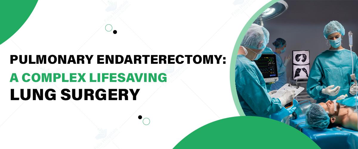
Pulmonary Endarterectomy (PEA) is a complex yet lifesaving surgical procedure for those suffering from chronic thromboembolic pulmonary hypertension (CTEPH). This surgery aims to remove blockages in the pulmonary arteries, improving blood flow and relieving symptoms such as shortness of breath and chest pain. For anyone considering this surgery, it's essential to choose the best hospital for Pulmonary Endarterectomy to ensure optimal outcomes and advanced post-operative care. Felix Hospital provides high-quality care with a team of experienced specialists, including Dr. Priyadarshi Jitendra Kumar, renowned for his expertise in pulmonology.
If you or a loved one is dealing with CTEPH, don’t hesitate to Click Here to reach out to Felix Hospital.
Pulmonary Endarterectomy (PEA) is a highly specialized surgical procedure used to treat CTEPH by removing organized blood clots from the pulmonary arteries. When blood clots from deep veins lodge in the pulmonary arteries, they obstruct blood flow and can lead to increased pressure in the lungs, resulting in CTEPH. PEA surgery removes these obstructions, allowing blood to flow freely, reducing lung pressure, and improving oxygenation.
Surgery is typically required in patients with CTEPH when:
1. Significant Blockage: Persistent blood clots obstruct the pulmonary arteries, causing increased pressure and symptoms.
2. Severe Symptoms: Patients experience severe shortness of breath, fatigue, or chest pain that impacts their quality of life.
3. Ineffectiveness of Medication: For some patients, medications are ineffective in managing CTEPH, making surgery the best option.
4. Risk of Heart Failure: Elevated lung pressure can lead to heart strain or right heart failure if untreated.
PEA is often a last-resort option, usually considered after confirming that other treatments or medications won’t suffice.
CTEPH primarily arises from unresolved pulmonary embolisms (PEs) or blood clots that form in the deep veins, usually in the legs, and travel to the lungs. When these clots do not dissolve, they can permanently lodge in the pulmonary arteries, causing long-term pressure and damage.
Other factors that may contribute to the development of CTEPH include:
Genetic predispositions
Chronic inflammatory or autoimmune conditions
Certain blood disorders
Previous surgeries or trauma
To accurately diagnose CTEPH and determine if PEA surgery is appropriate, a series of tests and imaging studies are performed:
1. Pulmonary Angiography: This imaging test provides detailed visuals of the pulmonary arteries to locate any obstructions.
2. Echocardiography: This test measures pressure within the pulmonary arteries and assesses heart function.
3. Ventilation-Perfusion (V/Q) Scan: A V/Q scan identifies regions in the lungs where blood flow may be restricted.
4. CT Pulmonary Angiography: CT angiography allows doctors to view blood flow within the pulmonary arteries and identify any blood clots.
5. Right Heart Catheterization: This procedure helps measure blood pressure in the lungs to confirm the presence of CTEPH.
The prognosis for patients undergoing PEA surgery is generally favorable, especially when treated at a well-equipped facility with expert surgeons. PEA provides substantial symptom relief, improved quality of life, and increased survival rates, though results can vary based on individual health factors. The cost of a Pulmonary Endarterectomy surgery depends on factors such as hospital facilities, surgeon fees, and post-operative care requirements. Patients are encouraged to discuss the expected costs with their healthcare provider to prepare for both the surgery and the recovery phase.
At Felix Hospital, we’re proud to have Dr. Priyadarshi Jitendra Kumar, one of the best pulmonologists, specializing in pulmonary surgeries and advanced respiratory care. Dr. Kumar’s extensive experience and patient-centered approach make him the ideal choice for anyone considering a PEA.
Pulmonary Endarterectomy is a challenging yet rewarding surgery that offers hope to those struggling with CTEPH. Choosing a hospital with the expertise, resources, and skilled pulmonologists can make a significant difference in your treatment outcome. At Felix Hospital, our team is here to provide comprehensive support through every stage of your journey to better lung health.
Ready to learn more about Pulmonary Endarterectomy and how we can help? Contact Felix Hospital today at +91 9667064100.
1. What is the main goal of Pulmonary Endarterectomy?
ANS: The main goal is to remove blood clots from the pulmonary arteries to restore normal blood flow and relieve symptoms of CTEPH.
2. Who qualifies for Pulmonary Endarterectomy?
ANS: Patients with CTEPH, where medications are ineffective, and the risk of heart failure is present, may be candidates.
3. How long is the recovery period?
ANS: Recovery varies, but most patients can resume normal activities within a few months, with gradual improvement over time.
4. What are the risks associated with PEA?
ANS: While generally safe, risks include bleeding, infection, or recurrence of symptoms, especially in patients with severe pre-existing conditions.
5. How effective is PEA in treating CTEPH?
ANS: PEA is highly effective, providing substantial symptom relief and improving the quality of life for many patients.
6. Is the procedure painful?
ANS: The surgery is performed under anesthesia, so patients do not experience pain during the procedure. Post-operative discomfort is managed with medications.
7. Does insurance cover the cost of Pulmonary Endarterectomy?
ANS: Many insurance plans cover the surgery, but coverage varies. It’s advisable to confirm with your provider.
8. Can lifestyle changes help after surgery?
ANS: Yes, lifestyle changes like regular exercise, a balanced diet, and avoiding smoking can enhance post-surgery recovery and overall health.
9. How do I prepare for PEA surgery?
ANS: Preparation includes medical evaluations, lifestyle adjustments, and following your doctor’s guidance on diet and medications leading up to surgery.

A COPD friendly diet is important for several reasons as discussed before. It can assist in relieving symptoms, controlling life threats and managing overweight.

For individuals seeking specialized care, a Pulmonary Edema Hospital in Noida offers advanced treatment options and expert management of this condition, ensuring comprehensive care for patients in need.
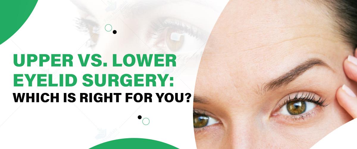
Eyelid surgery, also known as blepharoplasty, is a transformative procedure designed to rejuvenate the appearance of the eyes by addressing various concerns with the upper and lower eyelids. As one of the most popular cosmetic surgeries worldwide, eyelid surgery enhances both aesthetic appeal and functional performance. With age, the skin around the eyes tends to lose elasticity, leading to sagging, puffiness, and wrinkles that can make one look tired or older than they feel.
Understanding the differences between upper and lower eyelid surgery is crucial for making the right choice. Both procedures cater to distinct concerns and offer unique benefits. At a leading plastic surgery hospital, patients can benefit from expert guidance to determine the best procedure for their needs. This blog delves into the specifics of upper vs. lower eyelid surgeries to help you decide which one might be right for you.
Schedule a consultation with our expert surgeons today by Calling +919667064100 to explore your options and achieve your ideal look!
Upper eyelid surgery focuses on correcting issues related to the upper eyelids. It aims to restore a youthful and vibrant appearance by addressing functional and cosmetic concerns.
1. Excess Skin Causing Droopy Eyelids: Aging can lead to an accumulation of excess skin on the upper eyelids, resulting in a sagging appearance.
2. Impaired Vision Due to Sagging Eyelids: For some individuals, droopy upper eyelids can obstruct their field of vision.
3. Loss of Natural Eyelid Crease: Over time, the natural fold of the upper eyelid may become less defined.
The surgery involves the removal or repositioning of excess skin, fat, and sometimes muscle. Surgeons make precise incisions along the natural eyelid crease to minimize visible scarring.
The primary aesthetic goal is to restore a refreshed and youthful look, enhancing the overall harmony of facial features.
Improved peripheral vision.
Reduced strain on the eyes.
Lower eyelid surgery aims to enhance the appearance of the lower eye area, primarily addressing issues like under-eye bags, puffiness, and wrinkles.
1. Under-Eye Bags and Puffiness: These are often caused by fat deposits that become more prominent with age.
2. Dark Circles and Hollowing: Loss of volume and changes in skin tone can create a tired look.
3. Wrinkles and Sagging Skin Under the Eyes: Aging and reduced skin elasticity contribute to these concerns.
The procedure involves removing or redistributing fat, tightening skin, or both. Surgeons make incisions either inside the lower eyelid (transconjunctival approach) or just below the lash line to minimize scarring.
Lower eyelid surgery aims to create a smoother, rejuvenated under-eye area, reducing signs of fatigue and aging.
Reduced discomfort caused by excess skin or puffiness.
Enhanced self-confidence due to a more youthful appearance.
Targeted Areas: Upper eyelid surgery focuses on the area above the eyes, while lower eyelid surgery targets the area below the eyes.
Common Concerns Addressed: Upper eyelid surgery typically addresses drooping and vision impairment, whereas lower eyelid surgery focuses on puffiness, bags, and wrinkles.
Techniques and Surgical Approaches: The incision locations and techniques differ for each procedure.
Procedure Time: Both surgeries usually take 1-2 hours, depending on complexity.
Recovery Time: Recovery for upper eyelid surgery often ranges from 1-2 weeks, while lower eyelid surgery may take slightly longer due to swelling and bruising.
Upper eyelid scars are usually hidden within the natural crease, making them nearly invisible.
Lower eyelid scars, if any, are discreetly placed along the lash line or inside the eyelid.
Ideal candidates for upper eyelid surgery often experience drooping eyelids or vision issues.
Lower eyelid surgery candidates usually struggle with under-eye bags, puffiness, or wrinkles.
In some cases, patients may benefit from a combination of upper vs. lower eyelid surgery. This approach is particularly suitable for individuals with significant aging signs affecting both the upper and lower eyelids. Combining procedures can:
Enhance overall facial harmony.
Provide a comprehensive rejuvenation.
Reduce total recovery time compared to undergoing the surgeries separately.
Determining the most suitable procedure requires a personalized consultation with a board-certified plastic surgeon. During the consultation, the surgeon will:
Assess your concerns and goals.
Evaluate your facial anatomy and skin condition.
Recommend the procedure(s) that align with your needs.
For the best outcomes in eyelid surgery, trust Dr. Pragya Priya and Dr. Vishvendu Gaur, highly skilled and experienced plastic surgeons at Felix Hospital. Committed to excellence, Dr. Priya and Dr. Gaur provide personalized care to ensure optimal results. Whether you’re considering upper eyelid surgery, lower eyelid surgery, or a combination of both, they offer expert guidance to help you make informed decisions. At Felix Hospital, patients benefit from advanced facilities and a patient-focused approach, making it the preferred choice for eyelid surgery and other cosmetic procedures.
Click here to learn more about upper and lower eyelid surgery and discover the best solution for your needs.
Eyelid surgery is a powerful solution for addressing signs of aging, improving vision, and enhancing facial aesthetics. Whether you choose upper vs. lower eyelid surgery depends on your unique concerns and goals. For some, a combination of both may provide the best results. Understanding the differences between upper and lower eyelid surgery is essential in making the right choice. Consulting a qualified plastic surgeon at Felix Hospital, ensures you receive tailored advice and exceptional care. Take the first step toward brighter, refreshed eyes.
1. What is the recovery process like for upper eyelid surgery compared to lower eyelid surgery?
Ans: Recovery from upper eyelid surgery typically takes 1-2 weeks, with minimal swelling and bruising. Lower eyelid surgery may involve more swelling and take slightly longer to heal due to the delicate nature of the under-eye area.
2. Can upper and lower eyelid surgeries be performed at the same time?
Ans: Yes, combining both procedures is common for patients with concerns affecting both upper and lower eyelids. It provides a comprehensive rejuvenation and reduces overall recovery time.
3. Will eyelid surgery leave visible scars?
Ans: Upper eyelid scars are usually hidden in the natural crease of the eyelid, making them nearly invisible. For lower eyelid surgery, scars are either placed inside the eyelid (transconjunctival approach) or just below the lash line to remain discreet.
4. How do I know if I need upper or lower eyelid surgery?
Ans: Upper eyelid surgery is ideal for addressing droopy eyelids and vision impairment, while lower eyelid surgery targets under-eye bags, wrinkles, and puffiness. A consultation with a plastic surgeon can help determine the most appropriate procedure for your concerns.
5. What type of anesthesia is used during eyelid surgery?
Ans: Both upper and lower eyelid surgeries are typically performed under local anesthesia with sedation or general anesthesia, depending on the complexity of the procedure and patient preference.
6. Are the results of upper or lower eyelid surgery permanent?
Ans: While the results are long-lasting, aging will continue to affect the skin over time. Most patients enjoy the benefits of eyelid surgery for 10-15 years before considering touch-ups.
7. Can eyelid surgery correct dark circles under the eyes?
Ans: Lower eyelid surgery can address dark circles caused by under-eye bags or hollowing, but it may not fully eliminate discoloration due to pigmentation. Additional treatments like fillers or skin resurfacing may be recommended for optimal results.
8. Is eyelid surgery covered by insurance?
Ans: Insurance may cover upper eyelid surgery if drooping eyelids impair vision. Lower eyelid surgery is generally considered a cosmetic procedure and is unlikely to be covered. It's best to check with your provider for specific details.

Blocked arteries, or atherosclerosis, occur when plaque builds up in the arteries, restricting blood flow to the heart and increasing the risk of heart disease. Adopting a heart-healthy lifestyle and implementing expert-backed strategies can help clear arteries and improve cardiovascular health. This blog covers five key tips to reduce artery blockage, support heart health, and lower your risk of heart-related complications. For those seeking professional care, choosing the best heart hospital in Noida can also provide the guidance needed to keep your heart in top shape.
Reach out to a healthcare provider for personalized advice on maintaining clear arteries and a strong heart. Call us at +91 9667064100.
One of the most effective ways to reduce artery blockage is through a heart-healthy diet. Experts agree that the foods you eat directly impact cholesterol levels, blood pressure, and inflammation, which are all key factors in artery health.
Focus on Fiber: High-fiber foods, such as fruits, vegetables, whole grains, and legumes, can lower LDL (bad) cholesterol levels and help prevent plaque buildup.
Choose Healthy Fats: Opt for unsaturated fats, found in olive oil, avocados, and nuts, which can reduce bad cholesterol. Omega-3 fatty acids from fish like salmon, mackerel, and trout can also help decrease inflammation.
Avoid Trans Fats and Limit Saturated Fats: Trans fats, often found in processed foods, raise bad cholesterol while lowering good cholesterol, increasing plaque risk. Saturated fats, found in red meat and full-fat dairy, should be limited to maintain artery health.
Increase Antioxidants: Antioxidant-rich foods, such as berries, leafy greens, and tomatoes, help prevent oxidative stress, which contributes to artery damage and plaque buildup.
The American Heart Association (AHA) recommends adopting a Mediterranean or DASH (Dietary Approaches to Stop Hypertension) diet, both of which have been shown to reduce heart disease risk by lowering blood pressure and cholesterol.
Regular physical activity plays a crucial role in reducing artery blockage and improving heart health. Exercise helps lower blood pressure, improves cholesterol levels, and enhances circulation, which are all essential for maintaining clear arteries.
Aerobic Exercise: Activities like brisk walking, cycling, swimming, or running strengthen the heart and help clear arterial plaque. Aim for at least 150 minutes of moderate-intensity aerobic exercise per week.
Strength Training: Incorporating weight training into your routine twice a week can improve overall cardiovascular health and support weight management, a key factor in reducing heart disease risk.
Flexibility and Balance Exercises: Yoga and stretching not only reduce stress but also improve flexibility, which supports overall cardiovascular function.
According to the American College of Sports Medicine (ACSM), consistent exercise can reduce arterial stiffness, which can lower the risk of plaque buildup. Regular physical activity improves the endothelium’s function—the thin membrane inside blood vessels—preventing cholesterol from adhering to artery walls.
Chronic stress contributes significantly to heart disease risk. When you’re stressed, your body releases stress hormones like cortisol and adrenaline, which can increase blood pressure, promote inflammation, and make arterial plaque more likely to form. Managing stress is essential for keeping arteries clear and supporting heart health.
Practice Mindfulness and Meditation: Mindfulness practices, such as deep breathing and meditation, have been shown to lower blood pressure and reduce stress hormones. Even a few minutes daily can positively impact cardiovascular health.
Stay Socially Connected: Isolation can contribute to stress and anxiety, both of which affect heart health. Regularly connecting with friends, family, or support groups can improve mood and reduce stress.
Engage in Hobbies and Relaxation Activities: Reading, cooking, gardening, or any enjoyable hobby can help distract from stress and foster a sense of well-being, supporting a healthy heart.
The American Psychological Association (APA) notes that chronic stress can lead to inflammation and arterial damage, which increases plaque buildup. Stress management techniques like yoga, deep breathing, and social interaction have been proven to reduce these effects.
Smoking is one of the leading causes of arterial blockage, as it damages the lining of arteries, making it easier for plaque to accumulate. The chemicals in cigarette smoke increase blood pressure, reduce oxygen levels, and contribute to inflammation, all of which can cause arterial damage. Similarly, excessive alcohol consumption can raise blood pressure and triglyceride levels, increasing the risk of atherosclerosis.
Seek Support Programs: Many hospitals, clinics, and organizations offer programs to help individuals quit smoking, often through counseling and support groups.
Consider Nicotine Replacement Therapy (NRT): Options like patches, gum, and lozenges can reduce withdrawal symptoms and help break the addiction.
Try Medication: Certain medications, such as varenicline (Chantix) or bupropion (Zyban), are prescribed to help reduce cravings and ease the quitting process.
Moderation is Key: Limit alcohol intake to one drink per day for women and two for men. Red wine in moderation, for instance, has some heart-protective benefits, but excessive drinking poses a risk.
According to the Centers for Disease Control and Prevention (CDC), quitting smoking reduces the risk of heart disease within a few years of cessation, even for long-term smokers. Limiting alcohol can similarly lower the risk of high blood pressure and plaque buildup.
Several health conditions, including high blood pressure, diabetes, and high cholesterol, are directly linked to artery blockage and heart disease. Regular monitoring and proactive management of these conditions can significantly reduce the risk of arterial plaque buildup.
Control Blood Pressure: High blood pressure damages arteries over time, making it easier for plaque to form. Lifestyle changes, combined with medication if necessary, can help keep blood pressure in a healthy range.
Manage Blood Sugar Levels: High blood sugar levels, especially in those with diabetes, can damage blood vessels and increase the risk of plaque accumulation. A balanced diet, regular exercise, and medication, if needed, can help maintain stable blood sugar.
Lower Cholesterol Levels: High LDL cholesterol can lead to plaque buildup, while HDL cholesterol helps clear it from the arteries. Regular cholesterol checks and a heart-healthy diet can help maintain optimal levels.
The National Heart, Lung, and Blood Institute (NHLBI) recommends regular health screenings and working with healthcare providers to manage conditions like hypertension, diabetes, and high cholesterol, as these are among the most common contributors to arterial blockage.
In addition to these expert-backed strategies, here are some additional tips to further support heart health and reduce the risk of artery blockage:
Maintain a Healthy Weight: Excess body weight puts additional strain on the heart and increases the risk of developing atherosclerosis.
Stay Hydrated: Adequate hydration improves circulation and helps the body flush out toxins, supporting cardiovascular function.
Get Regular Sleep: Poor sleep can lead to elevated stress levels, high blood pressure, and inflammation, all of which can impact artery health.
Limit Sugar Intake: High sugar intake has been linked to increased risk of arterial plaque formation. Opt for whole foods and limit sugary snacks and beverages.
At Felix Hospital, our team of the best cardiologists in Noida is dedicated to helping you achieve and maintain a healthy heart. Our specialists include:
With their expertise, you'll receive a thorough evaluation and a personalized treatment plan focused on preventing artery blockage and boosting heart health.
Take a proactive step today—Click Here to schedule your heart health check-up and embark on your journey to better heart health!
Reducing artery blockage and boosting heart health involves a combination of diet, exercise, stress management, and lifestyle changes. By following these five expert-backed tips—adopting a heart-healthy diet, staying physically active, managing stress, avoiding smoking and limiting alcohol, and monitoring health conditions—you can significantly reduce the risk of plaque buildup and promote a healthier heart. Consistency is key; even small changes add up over time, making a big difference in long-term heart health. Take control today, and give your heart the care it deserves!
Q-What causes artery blockage?
ANS: Artery blockage, or atherosclerosis, is primarily caused by the buildup of plaque in the arteries. Plaque forms from cholesterol, fat, calcium, and other substances in the blood, leading to restricted blood flow over time.
Q- Can artery blockage be reversed naturally?
ANS: While completely reversing plaque buildup is challenging, lifestyle changes such as a healthy diet, exercise, and stress management can help reduce further blockage, improve blood flow, and support artery health.
Q- What foods should I avoid to prevent artery blockage?
ANS: Foods high in trans fats, saturated fats, and added sugars, such as processed foods, fried foods, sugary snacks, and red meat, can increase cholesterol levels and contribute to plaque formation.
Q- How does exercise help clear arteries?
ANS: Regular exercise can reduce cholesterol levels, improve circulation, and strengthen the heart. It also helps prevent high blood pressure, one of the main risk factors for artery blockage.
Q- Is stress linked to heart disease and blocked arteries?
ANS: Yes, chronic stress raises blood pressure, increases inflammation, and can lead to unhealthy behaviors, all of which elevate the risk of heart disease and artery blockage.
Q- How can I quit smoking to improve my heart health?
ANS: Many people find success with a combination of support groups, nicotine replacement therapy (like patches or gum), and counseling. A healthcare provider can help you find a quitting strategy tailored to your needs.
Q- What’s the best type of diet to support artery health?
ANS: The Mediterranean and DASH diets are both excellent options for heart health. They focus on fiber-rich foods, healthy fats, lean proteins, and antioxidant-rich fruits and vegetables, all of which help keep arteries clear.
Q- How often should I get my cholesterol and blood pressure checked?
ANS: For adults with no previous heart conditions, an annual checkup is recommended. If you have a history of high cholesterol or blood pressure, more frequent monitoring may be necessary.
Q- Are there any symptoms of early artery blockage?
ANS: Artery blockage often progresses without symptoms initially. However, over time, you may experience chest pain, shortness of breath, fatigue, or discomfort in the arms or neck. If you experience these, consult a doctor immediately.

हार्ट अटैक के मुख्य लक्षण, कारण और तुरंत किए जाने वाले उपचार के बारे में जानें। समय पर पहचान और सही इलाज से जीवन बचाया जा सकता है।
Arrhythmia is a condition where the heart beats irregularly—too fast, too slow, or with an uneven rhythm. It can feel like a fluttering, racing, or skipped heartbeat.
Laminectomy surgery is an effective treatment for various spine conditions, offering relief from pain, weakness, and mobility issues caused by spinal cord or nerve compression. If you or a loved one are considering this procedure at a neuro hospital in Noida, understanding the cost structure and factors that influence it is essential. This guide will break down the costs, and the factors that affect them, and provide insights on how to make informed decisions to ensure a successful surgery and recovery.
If you’re experiencing chronic back or leg pain, don’t wait! Call +91 9667064100 now.
Laminectomy is a surgical procedure performed to relieve pressure on the spinal cord or nerves. It involves the removal of the lamina, a bony structure that forms part of the vertebra, creating more space in the spinal canal and alleviating nerve compression. This surgery is commonly recommended for conditions like spinal stenosis, herniated discs, and other spinal issues that cause pain, numbness, and weakness in the legs and arms.
The cost of laminectomy surgery can vary based on various factors, such as the complexity of the case, the type of hospital, and additional medical needs. Here’s an approximate breakdown of the costs in Noida:
| Minimum Cost | 1,00,000 |
| Maximum Cost | 1,75,000 |
| Average Cost | 2,50,000 |
For more affordable and high-quality treatment options, Click Here to connect with the best hospital in Noida for laminectomy surgery today!
Several factors can influence the total cost of laminectomy surgery. Some of the key factors include:
Insurance Coverage: If you have insurance, it can cover a significant portion of the cost, reducing your out-of-pocket expenses.
While the cost of laminectomy surgery can be significant, here are a few tips to reduce the overall expense:
Felix Hospital, the best neuro hospital in Noida stands out as a premier choice for laminectomy surgery due to:
If you're considering laminectomy or disc surgery, it's crucial to work with a skilled neurosurgeon to ensure the best possible care. At our neuro hospital in Noida, we have a team of highly experienced surgeons who specialize in spine procedures, including:
Consulting these experts ensures that you receive high-quality care, helping you better understand the cost factors involved and determine the most suitable treatment plan for your condition.
Get in touch with us today to learn more about laminectomy surgery and the associated costs. Schedule your consultation by Clicking Here.
Laminectomy surgery is a highly effective procedure for alleviating chronic pain caused by spine-related conditions. While the cost can vary, understanding the factors involved and exploring ways to manage expenses can make the process more affordable. By choosing a trusted neuro hospital in Noida like Felix Hospital in Noida, you ensure that you receive the best care for both your health and your finances.
1. What is the average cost of laminectomy surgery in Noida, and how does it compare to other cities in India?
Ans: The cost of laminectomy surgery in Noida typically ranges between ₹1,50,000 to ₹2,50,000, depending on the hospital and the complexity of the procedure. It is often more affordable compared to metropolitan cities like Delhi and Mumbai, while maintaining high standards of care.
2. Does the cost of laminectomy surgery include pre-surgical diagnostics and post-surgical care?
Ans: Most hospitals provide packages that include pre-surgical diagnostic tests (like MRI or CT scans) and post-surgical follow-ups. However, these inclusions vary, and you should confirm the details with the hospital beforehand.
3. Are there additional charges for specialized equipment used during minimally invasive laminectomy?
Ans: Yes, if minimally invasive techniques are employed, additional charges may apply for specialized surgical instruments or technology such as endoscopic equipment.
4. How much do hospital room charges affect the total cost of laminectomy surgery?
Ans: Room charges can significantly impact the overall cost. For instance, opting for a general ward might reduce expenses, while private or deluxe rooms can increase the bill by ₹10,000 to ₹30,000 per day.
5. Does the surgeon’s expertise influence the cost of laminectomy surgery in Noida?
Ans: Yes, the cost can vary based on the surgeon’s experience and specialization. Highly experienced neurosurgeons may charge a premium for their expertise, adding ₹50,000 to ₹1,00,000 to the total cost.
6. Is laminectomy surgery covered under health insurance plans in Noida?
Ans: Many health insurance plans cover laminectomy surgery as it is considered medically necessary for conditions like spinal stenosis. However, the extent of coverage depends on your policy, including co-payments and exclusions.
7. Are there cost differences between government hospitals and private hospitals in Noida?
Ans: Yes, government hospitals generally charge significantly less than private hospitals. However, private hospitals often offer shorter waiting times, advanced technology, and more personalized care.
8. What are the common out-of-pocket expenses associated with laminectomy surgery in Noida?
Ans: Out-of-pocket expenses may include transportation, post-operative physiotherapy, medications, and any additional tests required after discharge. These costs can range from ₹10,000 to ₹50,000.

A concise guide explaining brain tumor surgery cost in Noida, including key factors, treatment options, and expert care.

Multiple sclerosis is a neurological disease that affects the brain and spinal cord.

Migraine relief includes advanced treatments and simple lifestyle changes to reduce pain and prevent attacks.
दांतों की देखभाल के प्रति जागरूकता होने के बावजूद, कई बार हमें ऐसे दांतों की समस्याओं का सामना करना पड़ता है जिनका इलाज केवल विशेषज्ञ ही कर सकते हैं। दांतों के गंभीर संक्रमण या क्षति के मामलों में रूट कैनाल उपचार (Root Canal Treatment) एक प्रभावी समाधान है। यह प्रक्रिया दांत को बचाने और दर्द से राहत दिलाने में मदद करती है, लेकिन इसके बारे में कई भ्रांतियाँ लोगों के बीच फैली हुई हैं जिसके चलते आप सर्वश्रेष्ठ दंत चिकित्सालय से संपर्क करें । इस ब्लॉग में हम रूट कैनाल उपचार से संबंधित मिथकों और सच्चाइयों पर चर्चा करेंगे ताकि आपको इससे जुड़ी सही जानकारी प्राप्त हो सके।
क्या आप दांतों से जुड़ी किसी समस्या का सामना कर रहे हैं? आज ही फ़ेलिक्स हॉस्पिटल के अनुभवी दंत चिकित्सकों से संपर्क करें +91 9667064100।
रूट कैनाल उपचार एक सामान्य दंत चिकित्सा प्रक्रिया है जिसका उपयोग तब किया जाता है जब दांत की जड़ या पल्प (रक्त वाहिकाएं और तंत्रिकाएं) संक्रमित या क्षतिग्रस्त हो जाती है। इस उपचार में दांत के अंदर के संक्रमित पल्प को हटा दिया जाता है, और फिर उसे साफ करके एक विशेष भराव सामग्री से भर दिया जाता है ताकि दांत को भविष्य में किसी भी प्रकार के संक्रमण से बचाया जा सके।
रूट कैनाल प्रक्रिया का मुख्य उद्देश्य दांत को हटाने की बजाय उसे संरक्षित रखना होता है, जिससे मरीज की प्राकृतिक मुस्कान और चबाने की क्षमता को बनाए रखा जा सके।
रूट कैनाल उपचार को लेकर कई भ्रांतियाँ हैं, जो अक्सर लोगों को भ्रमित कर देती हैं। आइए जानते हैं कुछ सामान्य मिथकों के बारे में:
1. मिथक: रूट कैनाल बेहद दर्दनाक होता है।
सच्चाई: यह सबसे सामान्य मिथक है। लोग सोचते हैं कि रूट कैनाल उपचार दर्द से भरा होता है, जबकि सच्चाई यह है कि यह उपचार आधुनिक तकनीकों और एनेस्थीसिया (निश्चेतक) के उपयोग से लगभग दर्द रहित होता है। यह उपचार दर्द को खत्म करने के लिए किया जाता है, न कि इसे बढ़ाने के लिए।
2. मिथक: रूट कैनाल के बाद दांत कमजोर हो जाता है।
सच्चाई: रूट कैनाल के बाद दांत कमजोर नहीं होता, बल्कि उसे सही तरीके से संरक्षित किया जाता है। अगर दांत में उचित क्राउन या कैप लगाया जाए, तो यह बिल्कुल सामान्य तरीके से कार्य करता है और कोई कमजोरी महसूस नहीं होती।
3. मिथक: रूट कैनाल इलाज के बाद दांत में संक्रमण फिर से हो सकता है।
सच्चाई: जब रूट कैनाल उपचार सही तरीके से किया जाता है और दांत को सील कर दिया जाता है, तो दोबारा संक्रमण होने की संभावना बहुत कम हो जाती है। हालांकि, नियमित दंत देखभाल और साफ-सफाई बनाए रखना आवश्यक है।
4. मिथक: रूट कैनाल एक महंगा उपचार है।
सच्चाई: रूट कैनाल की कीमत उस पर निर्भर करती है कि आपको किस प्रकार के दांत में समस्या है और उपचार कितना जटिल है। इसके अलावा, यदि इसे न करवाया जाए तो दांत हटाने और प्रत्यारोपण जैसी अन्य प्रक्रियाएं और भी महंगी हो सकती हैं।
5. मिथक: रूट कैनाल के बजाय दांत निकालना बेहतर है।
सच्चाई: रूट कैनाल का मुख्य उद्देश्य दांत को बचाना है। एक बार दांत निकाल दिया जाए, तो उसकी जगह कृत्रिम दांत लगाना ही विकल्प बचता है, जो कि अधिक महंगा और जटिल होता है। प्राकृतिक दांत की जगह कुछ भी नहीं ले सकता, इसलिए दांत को बचाना हमेशा प्राथमिकता होनी चाहिए।
अब जब हमने मिथकों पर चर्चा कर ली है, आइए रूट कैनाल से जुड़ी कुछ महत्वपूर्ण सच्चाइयों पर ध्यान दें:
1. दांत को संरक्षित करने का एकमात्र उपाय
रूट कैनाल उपचार दांत को निकालने से बचाने का सबसे प्रभावी तरीका है। इसके माध्यम से दांत के अंदर के संक्रमित हिस्से को हटा दिया जाता है और दांत को फिर से स्वस्थ बनाने के लिए सील किया जाता है।
2. दर्द से राहत
रूट कैनाल उपचार का मुख्य उद्देश्य दांत दर्द से छुटकारा दिलाना है। जब दांत की जड़ में संक्रमण होता है, तो यह असहनीय दर्द का कारण बन सकता है, जिसे रूट कैनाल के जरिए ठीक किया जा सकता है।
3. लंबी अवधि तक दांत को संरक्षित करता है
एक सफल रूट कैनाल उपचार के बाद, दांत वर्षों तक कार्य कर सकता है, बशर्ते उसकी उचित देखभाल की जाए। इससे मरीज की चबाने की क्षमता बनी रहती है और दांत की सुंदरता भी बरकरार रहती है।
4. उपचार की सफलता दर
रूट कैनाल की सफलता दर काफी उच्च होती है। जब यह उपचार विशेषज्ञ द्वारा सही ढंग से किया जाता है, तो इसका परिणाम दीर्घकालिक और प्रभावी होता है।
5. उपचार के बाद देखभाल की आवश्यकता
रूट कैनाल के बाद उचित देखभाल और दांतों की सफाई बेहद जरूरी है। नियमित ब्रशिंग, फ्लॉसिंग और दंत चिकित्सक के पास समय-समय पर चेकअप कराना आवश्यक है ताकि दांत स्वस्थ बना रहे।
नोएडा में रूट कैनाल ट्रीटमेंट (आरसीटी) हॉस्पिटल, फ़ेलिक्स हॉस्पिटल में दंत चिकित्सा के विशेषज्ञ अत्याधुनिक तकनीकों और उपकरणों का उपयोग करते हैं ताकि आपको सर्वोत्तम रूट कैनाल उपचार प्राप्त हो सके। हमारे अनुभवी दंत चिकित्सक यह सुनिश्चित करते हैं कि आपका उपचार दर्द रहित हो और आपको सर्वोत्तम परिणाम मिले। फ़ेलिक्स हॉस्पिटल में रूट कैनाल उपचार की विशेषताएँ:
फेलिक्स हॉस्पिटल्स में हमारे अनुभवी दंत चिकित्सक आपको सबसे बेहतर दंत चिकित्सा सेवाएँ प्रदान करने के लिए तत्पर हैं। हमारे विशेषज्ञ न केवल आपकी दांतों की समस्याओं का समाधान करते हैं, बल्कि आपको सही देखभाल के लिए मार्गदर्शन भी प्रदान करते हैं।
अपनी दांतों की समस्याओं को नजरअंदाज न करें। यहां क्लिक करें और आज ही फ़ेलिक्स हॉस्पिटल के दंत विशेषज्ञों से संपर्क करें और अपने दांतों का बेहतरीन इलाज प्राप्त करें।
रूट कैनाल उपचार एक प्रभावी और सुरक्षित तरीका है जिससे दांतों के संक्रमण को ठीक किया जा सकता है और दांत को संरक्षित रखा जा सकता है। हालाँकि इसके बारे में कई मिथक प्रचलित हैं, लेकिन वास्तविकता यह है कि यह उपचार न केवल दर्द से राहत दिलाता है बल्कि लंबे समय तक दांतों को स्वस्थ भी रखता है। सही समय पर उपचार कराने से आप दांत निकालने से बच सकते हैं और अपनी प्राकृतिक मुस्कान को बनाए रख सकते हैं।
1. क्या रूट कैनाल उपचार दर्दनाक होता है?
उत्तर: नहीं, आधुनिक एनेस्थीसिया और तकनीकों के उपयोग से रूट कैनाल उपचार लगभग दर्द रहित होता है। प्रक्रिया के दौरान आपको दर्द महसूस नहीं होगा, और यह उपचार वास्तव में दर्द को कम करने के लिए किया जाता है।
2. रूट कैनाल उपचार कितने सत्रों में पूरा होता है?
उत्तर: आमतौर पर, रूट कैनाल उपचार 1-2 सत्रों में पूरा हो सकता है, लेकिन यह दांत की स्थिति और संक्रमण की गंभीरता पर निर्भर करता है। कुछ मामलों में, 3 सत्रों की भी आवश्यकता हो सकती है।
3. क्या रूट कैनाल के बाद दांत में कमजोरी आती है?
उत्तर: रूट कैनाल के बाद दांत को मजबूत बनाने के लिए उस पर क्राउन लगाया जाता है। यदि क्राउन सही तरीके से लगाया जाए, तो दांत सामान्य रूप से काम करता है और उसमें कमजोरी नहीं आती।
4. रूट कैनाल के बाद कितने समय तक दांत स्वस्थ रहता है?
उत्तर: रूट कैनाल के बाद यदि सही देखभाल की जाए, तो दांत लंबे समय तक स्वस्थ रहता है। नियमित रूप से दंत चिकित्सक से चेकअप और दांतों की सफाई करना आवश्यक होता है।
5. रूट कैनाल उपचार के बाद क्या किसी विशेष आहार की आवश्यकता होती है?
उत्तर: उपचार के बाद कुछ समय के लिए सख्त या बहुत गर्म भोजन से बचने की सलाह दी जाती है। दांत पर क्राउन लगने के बाद आप सामान्य आहार ले सकते हैं।
6. क्या रूट कैनाल के बाद संक्रमण दोबारा हो सकता है?
उत्तर: यदि उपचार सही तरीके से किया जाए और दांत की उचित देखभाल की जाए, तो दोबारा संक्रमण होने की संभावना कम होती है। नियमित दंत सफाई और चेकअप आवश्यक होते हैं।
7. क्या रूट कैनाल उपचार महंगा होता है?
उत्तर: रूट कैनाल उपचार की कीमत संक्रमण की गंभीरता और दांत की स्थिति पर निर्भर करती है। यह दांत निकालने और कृत्रिम दांत लगाने से अधिक किफायती हो सकता है।
8. रूट कैनाल की आवश्यकता कैसे पता चलेगी?
उत्तर: यदि आपको दांत में गंभीर दर्द, सूजन, या ठंडे या गर्म खाने पर अत्यधिक संवेदनशीलता महसूस हो रही है, तो यह संकेत हो सकता है कि आपको रूट कैनाल की आवश्यकता है। फ़ेलिक्स हॉस्पिटल के दंत चिकित्सक से परामर्श कराएं।
मसूड़ों से खून आना दांतों और मसूड़ों की समस्या का संकेत हो सकता है। जानिए इसके कारण, बचाव और प्रभावी इलाज के तरीके।
मसूड़ों की बीमारियाँ, जैसे कि पेरियोडोन्टल रोग (Periodontal Disease) या गम डिज़ीज़, एक आम समस्या है जो मसूड़ों के स्वास्थ्य को प्रभावित कर सकती है।
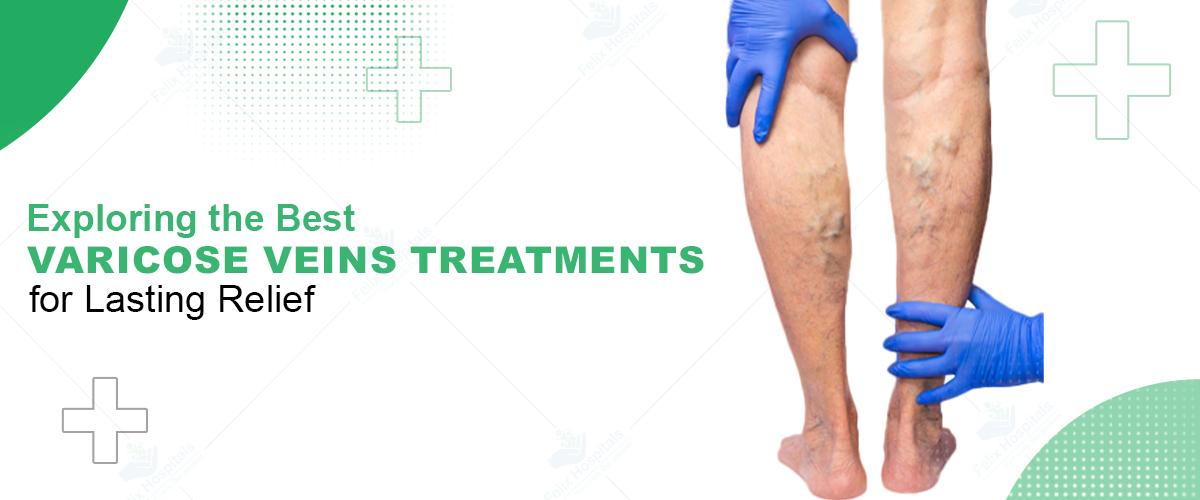
Varicose veins are enlarged, swollen ones often visible beneath the skin's surface, primarily affecting the legs, feet, or ankles. They occur when vein walls weaken and valves in the veins malfunction, leading to blood pooling and creating bluish or purple bulges. While many people view varicose veins as a cosmetic issue, they can cause discomfort and, in severe cases, complications. If you're experiencing symptoms, consult a specialist at a leading vascular surgery hospital in Noida for guidance on tailored treatment options.
Contact Felix Hospital today for your good health tomorrow. Call us at +91 9667064100.
Varicose veins are bulging blood vessels visible through the skin, primarily in the legs. They range in color from blue to purple and often have a twisted, cord-like appearance. Symptoms can include pain, itching, or a burning sensation, though the condition is generally not dangerous. However, in severe cases, complications such as blood clots may develop. While home care can ease symptoms, more advanced cases may require medical treatments like injections, laser therapy, or surgery.
Visible symptoms include blue or purple veins that appear twisted and bulging. Although varicose veins are often painless, they can lead to:
Aching or heaviness in the legs
Burning, throbbing, muscle cramps, and swelling
Increased pain after prolonged sitting or standing
Skin discoloration around the affected veins
Smaller veins, known as spider veins, may also appear close to the skin's surface and vary in size and color. They commonly appear on the face and legs.
Top Causes Varicose veins Treatment develop when vein walls weaken, allowing them to stretch under increased blood pressure. As veins expand, the valves within them—responsible for keeping blood flowing in one direction—start to fail. This failure causes blood to flow backward, leading it to pool within the vein, which swells, twists, and bulges as a result.
Natural aging.
Long periods of standing.
Hormonal changes.
Excess body weight.
Diagnosis typically involves a physical examination and a venous Doppler ultrasound to assess blood flow and check for blood clots in the veins.
Treatment ranges from self-care to surgical procedures, with various approaches to managing discomfort and improving blood flow:
Self-Care: Simple practices like elevating your legs, exercising, and wearing compression stockings can alleviate symptoms.
Compression Stockings: These assist veins and leg muscles in circulating blood. Prescription-strength compression stockings may be covered by insurance if there are symptoms.
Procedures and Surgeries: If symptoms persist or varicose veins are severe, options include:
Sclerotherapy: A solution is injected to close off the veins.
Laser Treatment: Light bursts target veins, causing them to fade.
Catheter Procedures: Radiofrequency or laser energy collapses large veins.
Vein Stripping and Ligation: A surgical procedure where varicose veins are tied off and removed.
Ambulatory Phlebectomy: Small veins are removed through tiny skin pricks, often as an outpatient procedure.
The Cost of vascular vein treatment in Noida varies based on several factors, including the specific location, type of treatment, and facilities available.
Certain lifestyle changes can be beneficial in managing varicose vein symptoms and preventing their worsening:
Exercise: Walking helps improve blood circulation.
Weight Management: Reducing weight lessens the pressure on veins.
Dietary Adjustments: Limiting salt intake can reduce water retention, alleviating swelling.
Clothing Choices: Avoid tight clothes, as they restrict blood flow.
Leg Elevation: Raising the legs improves blood circulation and reduces pressure on veins.
Frequent Position Changes: Standing or sitting for extended periods restricts blood flow, so shifting positions regularly is helpful.
Family History: Genetics can increase the risk of developing varicose veins.
Obesity: Extra weight puts additional pressure on the veins.
Other Factors: Aging, hormonal changes (especially during pregnancy), and prolonged standing or sitting contribute to varicose vein development.
While rare, varicose veins can lead to certain complications:
Ulcers: Painful sores can form near varicose veins, particularly around the ankles.
Blood Clots: Varicose veins can be associated with blood clots, leading to leg pain and swelling.
Bleeding: In rare cases, veins close to the skin's surface may burst, causing minor bleeding.
Leg Swelling: Long-standing varicose veins can contribute to persistent swelling in the legs.
To minimize the risk of varicose veins or prevent them from worsening, adopt the following habits:
Limit High Heels and Tight Stockings: These can restrict circulation.
Stay Active: Frequent movement and exercise support healthy blood flow.
Balanced Diet: A diet high in fiber and low in salt helps reduce swelling.
Leg Elevation and Movement: Regularly elevating legs and moving throughout the day prevent blood from pooling.
For those seeking effective treatment for varicose veins, Dr. Ritesh Agrawal and Dr. Shruti Sharma recognized as the top vascular surgeon in Noida, is a highly experienced specialist at Felix Hospital. Committed to providing personalized care, he uses advanced techniques to ensure optimal outcomes for patients. Schedule a consultation today to discuss your symptoms and explore the best treatment options for you.
Book an appointment with Dr. Ritesh Agrawal , Call us at +91 9667064100 and taking the first step toward healthier veins!
Varicose veins may not always pose a serious health risk, but they can lead to discomfort and complications if left untreated. Understanding the causes, symptoms, and treatment options is crucial for managing this condition effectively. If you are experiencing symptoms, don't hesitate to reach out to a specialist for evaluation and care.
Q.1-What are the primary symptoms of varicose veins?
Ans: Common symptoms include visible blue or purple veins, aching or heaviness in the legs, swelling, muscle cramps, and a burning sensation, especially after long periods of sitting or standing.
Q.2-Can varicose veins go away on their own?
Ans: While mild cases may improve with lifestyle changes, varicose veins typically do not resolve on their own and may require medical treatment if symptoms persist or worsen.
Q.3- Are varicose veins a sign of a serious health condition?
Ans: While varicose veins are often a cosmetic concern, they can be associated with complications such as blood clots or skin ulcers if left untreated.
Q. 4- What lifestyle changes can help manage varicose veins?
Ans: Maintaining a healthy weight, exercising regularly, elevating the legs, wearing compression stockings, and avoiding prolonged periods of standing or sitting can help manage symptoms.
Q.5- Is there a genetic predisposition to developing varicose veins?
Ans: Yes, a family history of varicose veins can increase your likelihood of developing the condition.
Q.6- How do compression stockings work for varicose veins?
Ans: Compression stockings apply pressure to the legs, helping veins and leg muscles to circulate blood more effectively and reduce swelling and discomfort.
Q.7- What types of medical treatments are available for varicose veins?
Ans: Treatments include sclerotherapy, laser therapy, catheter procedures, vein stripping, and ambulatory phlebectomy, among others.
Q.8- Can pregnancy cause varicose veins?
Ans: Yes, hormonal changes and increased blood volume during pregnancy can lead to the development or worsening of varicose veins.
Q.9- How can I differentiate between varicose veins and spider veins?
Ans : Varicose veins are larger, bulging veins that often cause discomfort, while spider veins are smaller, flat, and typically do not cause symptoms.
A complete guide to understanding varicose vein surgery expenses in Noida, including treatment types, hospital charges, and recovery costs.
Meet Dr. Ritesh Agarwal, a leading specialist in varicose vein care. With extensive experience and a commitment to personalized treatment.

While varicose veins are often a cosmetic concern, they can also lead to symptoms like pain, discomfort, and even complications like ulcers or blood clots in severe cases.
Symptoms can include pain, itching, or a burning sensation, though the condition is generally not dangerous.

Subscribe to our
© Copyright 2026. All Rights Reserved by Felix Healthcare Private Limited
Design and development by :


 +(91)9667064100
 Emergency : +(91)9667064100