 WhatsApp

Subscribe to our
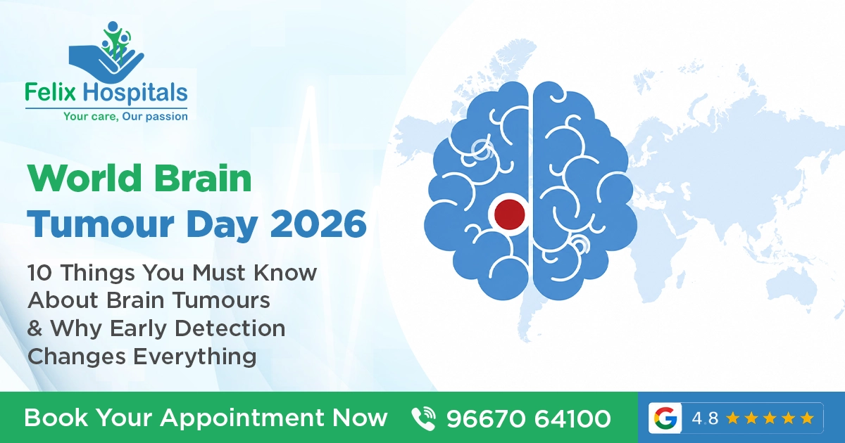
Every year on June 8, the world comes together to observe World Brain Tumour Day a day first established in 2000 by the German Brain Tumour Association (Deutsche Hirntumorhilfe e.V.) to raise awareness, reduce stigma, and push for better research, diagnosis, and treatment of brain tumours globally.
It is not a day built on fear. It is built on knowledge because when it comes to brain tumours, what you do not know can genuinely cost you your life.
At Felix Hospitals, we see the difference that early detection makes. We also see what happens when people ignore symptoms for months or years because they did not recognise what their body was trying to tell them.
This World Brain Tumour Day 2026, here are 10 things every person should know ranked by what matters most..
This is perhaps the most important thing to understand, and it changes how people think about the diagnosis entirely.
Brain tumours are broadly divided into two categories benign (non-cancerous) and malignant (cancerous). Benign tumours do not spread to other parts of the body. But and this is critical they can still be life-threatening depending on where they are located in the brain.
A benign tumour pressing against the part of the brain that controls breathing, vision, or movement can be just as dangerous as a malignant one. The brain has no extra space. There is no room to expand.
So when someone says "it is not cancer, you are fine," that is not the complete picture. What matters is location, size, rate of growth, and how quickly the right medical team gets involved.
According to global data, approximately 300,000 people are diagnosed with a primary brain tumour every year worldwide. In India, the incidence is estimated at around 5 to 10 per 100,000 population and with a population of 1.4 billion, that translates to a significant number of diagnoses annually.
What makes this particularly important is that brain tumours affect people of all ages from newborns to the elderly. They are, in fact, one of the most common cancers in children. In adults, the incidence increases with age, with the highest rates seen in people above 65.
The fact that brain tumours do not discriminate by age, gender, or lifestyle is exactly why awareness matters and why no persistent neurological symptom should be dismissed as stress or exhaustion without proper evaluation.
Brain tumours are deceptive. Many of their early symptoms overlap with far more common, benign conditions: tension headaches, vision problems from screen fatigue, mild personality changes written off as stress. This overlap is precisely why so many people are diagnosed late.
Here are the symptoms that warrant urgent medical evaluation:
None of these symptoms automatically mean a brain tumour. But any of them especially if persistent or progressive should be evaluated by a neurologist without delay.
Not all brain tumours are the same, and treatment is never one-size-fits-all. The World Health Organisation classifies brain tumours into more than 120 distinct types, based on the cell of origin, grade, and molecular characteristics.
Some of the most commonly encountered include:
Each type responds differently to surgery, radiation, and chemotherapy. This is why the histopathological and molecular diagnosis of the tumour is as important as identifying it on a scan.
An MRI showing a suspicious mass in the brain is not the end of the diagnostic process it is the beginning. Here is what typically follows:
Step 1 MRI with contrast: A gadolinium-enhanced MRI gives the treating team detailed information about the tumour's size, location, blood supply, and relationship to surrounding brain structures. Functional MRI (fMRI) may also be used to map critical brain regions before surgery.
Step 2 CT scan: Often used alongside MRI to assess bone involvement or detect calcification within the tumour.
Step 3 Biopsy or surgical resection: A tissue sample is essential for definitive diagnosis. This is analysed by a neuropathologist to determine the tumour type, grade, and increasingly, molecular markers that guide treatment decisions.
Step 4 Molecular profiling: Modern neuro-oncology relies heavily on molecular markers IDH mutation status, MGMT methylation, 1p/19q co-deletion, and others. These not only confirm diagnosis but also predict prognosis and response to specific therapies.
Step 5 Multidisciplinary team (MDT) review: Neurosurgeons, neuro-oncologists, radiation oncologists, radiologists, and neuropathologists review the case together before a treatment plan is finalised.
At Felix Hospital, this multidisciplinary approach is core to how complex neurological cases are managed. Every patient presenting with a suspicious neurological finding is evaluated through a structured protocol not a rushed, single-doctor decision.
Brain tumour treatment has evolved significantly over the last two decades. What was once limited to surgery and radiation now includes a range of modalities that can be combined and tailored to the specific tumour type and patient condition.
Surgery (Craniotomy): For many tumours, surgical removal or as much removal as safely possible remains the first line of treatment. Advances in neuronavigation, intraoperative MRI, and awake craniotomy (where the patient is kept conscious to monitor speech and motor function during surgery) have made resections safer than ever.
Radiation therapy: Used after surgery to target remaining tumour cells, or as primary treatment where surgery is not possible. Techniques include whole-brain radiotherapy, stereotactic radiosurgery (Gamma Knife, CyberKnife), and fractionated stereotactic radiotherapy.
Chemotherapy: Temozolomide (TMZ) is the most commonly used chemotherapy for high-grade gliomas. It is often given alongside and after radiation therapy.
Targeted therapy: For tumours with specific molecular profiles, targeted agents that block particular signalling pathways are now part of the treatment landscape.
Immunotherapy: Still largely investigational for brain tumours, but showing promise in certain subtypes, particularly glioblastoma.
Tumour Treating Fields (TTF): A wearable device therapy that uses low-intensity alternating electric fields to disrupt tumour cell division now approved for glioblastoma.
Rehabilitation: An often underappreciated component of brain tumour care. Physiotherapy, speech therapy, occupational therapy, and neuropsychological support are critical for recovery and quality of life after treatment.
One of the most distressing aspects of a brain tumour diagnosis for patients and families alike is the question: Why did this happen?
In the majority of cases, there is no clear identifiable cause. Unlike lung cancer and smoking, or cervical cancer and HPV, most brain tumours arise without a single definitive risk factor.
What we do know:
What the evidence does not support:
Understanding what is evidence-based and what is not matters because misinformation around brain tumours can lead people to make decisions based on fear rather than facts.
Brain tumours are the most common solid tumour in children and the leading cause of cancer-related death in the paediatric population. In children, the tumour types, locations, and treatment approaches differ significantly from adults.
Common paediatric brain tumours include:
Symptoms in children can be subtle and easily attributed to other causes headaches dismissed as school stress, balance problems written off as clumsiness, declining school performance seen as behavioural rather than neurological.
Parents and teachers should be aware: a child who suddenly develops coordination problems, complains of persistent morning headaches, or shows a rapid decline in academic performance or vision deserves neurological evaluation not just reassurance.
A brain tumour does not just affect the brain physically. It reshapes the lived experience of the person carrying it and their family.
Patients frequently report anxiety about recurrence, depression, cognitive changes (often called "chemo brain" or treatment-related cognitive impairment), and a profound sense of uncertainty about the future. Caregivers face their own invisible burden the emotional exhaustion of supporting someone through a diagnosis and treatment that can stretch over months or years.
Mental health support is not an optional add-on to brain tumour care. It is a core component.
This includes:
At Felix Hospital, every serious neurological diagnosis is approached with an understanding that the person in front of us is not just a scan or a biopsy result. They are someone whose life has just changed and who needs medical excellence and human support in equal measure.
World Brain Tumour Day is not just about awareness for others. It is an invitation to act.
If you have symptoms, get evaluated. Do not self-diagnose with Google. Do not wait for symptoms to worsen. An MRI takes less than an hour. The information it gives your doctor is irreplaceable.
If someone you know has symptoms, say something. People often dismiss their own neurological symptoms. A gentle, concerned push from someone who cares can be the reason they go to a doctor.
If you have a family history of brain tumours, discuss genetic counselling. Your personal risk assessment is worth having.
If you are a caregiver for someone with a brain tumour ask for help. For yourself. Caregiver burnout is real and well-documented. You cannot support someone else if you are running on empty.
If you want to contribute, support brain tumour research and advocacy. Patient advocacy organisations, research funding bodies, and hospitals working on trial therapies depend on public engagement and support.
Felix Hospital, is equipped to evaluate, diagnose, and manage patients presenting with neurological symptoms including those who may have a brain tumour.
Our neurology and neurosurgery team works within a multidisciplinary framework, ensuring that every patient receives a thorough evaluation before any treatment decision is made. Our diagnostic capabilities include advanced MRI, CT imaging, and access to neuropathological evaluation with a treatment approach that integrates surgery, oncology, rehabilitation, and psychological support.
If you or someone in your family is experiencing persistent neurological symptoms, headaches that are getting worse, new-onset seizures, unexplained vision or speech changes, progressive weakness do not delay.
Call Felix Hospital at +91 9667064100 to speak with our team or to schedule a neurological consultation.
No, The overwhelming majority of headaches are tension-type or migraine headaches, completely unrelated to brain tumours. Brain tumour headaches are typically progressive, worse in the morning, and accompanied by other neurological symptoms. A headache alone, even a severe one is rarely the only symptom of a brain tumour.
An MRI with contrast is the gold standard for brain tumour detection. A CT scan can pick up large lesions but is less sensitive than MRI. There is no blood test that reliably detects brain tumours. If a brain tumour is suspected clinically, imaging is non-negotiable.
Most brain tumours are not inherited. A small proportion are associated with hereditary genetic syndromes (such as neurofibromatosis). Having one family member with a brain tumour does not mean others will develop one, though genetic counselling may be appropriate in families with multiple affected individuals.
This depends heavily on the tumour type, grade, and location. Many benign tumours such as meningiomas and acoustic neuromas can be completely removed with an excellent prognosis. Some malignant tumours, particularly low-grade gliomas in adults, can be managed over many years with treatment. High-grade glioblastoma remains one of the most challenging cancers to treat, though research is actively advancing.
A primary brain tumour originates in the brain. A secondary (metastatic) brain tumour originates elsewhere in the body commonly the lung, breast, kidney, or skin and spreads to the brain. Secondary tumours are actually more common than primary brain tumours. Treatment differs significantly between the two.
Call +91 9667064100.Walk-ins are welcome for general queries, and appointments can be scheduled for specialist consultations.
Spot early brain tumor signs headaches, vision changes and see a neurologist in Noida quickly.
A blood clot in the brain can lead to serious conditions like stroke or brain hemorrhage. Understanding its causes, symptoms, and timely treatment is crucial for recovery.

Discover how Brain Angiograms in Noida are revolutionizing the diagnosis and treatment of neurological conditions with advanced imaging technology and expert care.
TBI affects brain function after injury; Felix Hospital, Noida provides expert care and recovery support.

Subscribe to our
© Copyright 2026. All Rights Reserved by Felix Healthcare Private Limited
Design and development by :


 +(91)9667064100
 Emergency : +(91)9667064100