 info@felixhospital.com
 +(91) 9667064100
 WhatsApp


Subscribe to our
Experiencing a blood clot in brain is scary and life-changing for many people every year. It can happen suddenly, leaving you feeling scared and helpless. But it's important to know that getting medical help right away can make a big difference. In this article, we'll talk about what causes of blood clot in brain, their symptoms, how doctors diagnose them, and the different treatments available.
Book Your Appointment One click at +91 9667064100
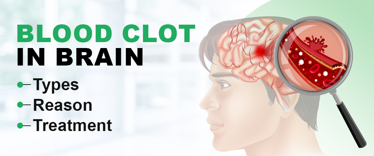
A blood clot in brain, also known as a stroke, is a serious emergency that can cause severe brain damage or even death. It happens when blood cells stick together and block a blood vessel in the brain. This stops oxygen and nutrients from getting to the brain, which can harm or kill brain cells. Let's go through the types, reasons, signs, diagnosis, and treatments for blood clots in the brain in simpler terms. When a blood vessel in the brain ruptures and becomes filled with blood, it creates a blockage in an artery, leading to a blood clot. This blockage can result in the brain being deprived of oxygen, causing damage to brain cells and potentially leading to death.
For those seeking top neurological care, Felix Hospital is widely regarded as the best neurology hospital in Noida, known for treating complex brain conditions like blood clots.
While some individuals may experience mild episodes of this occurrence without noticeable symptoms, others may develop severe complications such as seizures or fatality. However, even before these complications arise, there are warning signs to watch for. Recognizing these symptoms (symptoms for blood clot in head) is crucial, prompting timely treatment for blood clots in the brain.
Blood clots typically form as a natural response to injury, aiming to stop blood loss. However, when a blood clot obstructs blood flow within blood vessels, it poses significant danger. Two primary types of blood clots exist: thrombosis and embolism.
Cervical cancer originates from the growth of cells in the cervix, the lower part of the uterus connected to the vagina. Most cases of cervical cancer are linked to various strains of the human papillomavirus (HPV Vaccine), a common infection transmitted through sexual contact. While the body's immune system can often combat HPV Vaccine (cervical cancer vaccine) upon exposure, in a small percentage of individuals, the virus persists for an extended period, contributing to the development of cancerous cervical cells.
To minimize the risk of cervical cancer, individuals are advised to undergo screening tests and receive a cervical cancer vaccine designed to protect against HPV infection. In cases where cervical cancer is diagnosed, initial treatment typically involves surgery to remove the cancer. Additional treatment options may include medications to eliminate cancer cells, such as chemotherapy and targeted therapy. Radiation therapy, utilizing potent energy beams, may also be employed, sometimes in combination with low-dose chemotherapy.
The price for thrombectomy surgery (the removal of a blood clot) in India varies from Rs. 100,000 to Rs. 2,50,000. However, this cost range is somewhat arbitrary and contingent on a variety of factors, encompassing both patient-specific and treatment-related considerations.
Patient-specific factors that may influence the cost of thrombectomy surgery include the cause and nature of the blockage, the specific blood vessel affected, and the overall health condition of the patient. Conversely, treatment-related factors affecting the cost encompass the type of surgical procedure undertaken, the anesthesia administered, and the professional fees charged by the surgeon, among others.
For a more precise estimate of the cost tailored to your individual circumstances, it is advisable to schedule a complimentary consultation with top vascular surgeons in your vicinity. This consultation can facilitate a comprehensive evaluation and diagnosis, enabling you to obtain a more accurate understanding of the anticipated expenses associated with the surgery.
For inquiries about the cost of brain clot surgery at Felix Hospital, Call Now at +91 9667064100.
Ischemic stroke, the most prevalent type, constitutes a significant majority of all strokes. It manifests in two forms: thrombotic and embolic. Thrombotic stroke arises when a blood clot, or thrombus, obstructs a brain artery, impeding blood flow. Embolic stroke occurs when plaque or thrombus fragments relocate from their origin, obstructing a downstream artery—a situation termed an embolus. The extent of brain damage hinges on the precise location of the blockage within the artery.
Typically, the carotid or vertebral arteries remain partially unobstructed, allowing a small blood flow to reach the brain. However, this diminished flow deprives brain cells of nutrients, precipitating cellular dysfunction and stroke symptoms. Within a stroke, a core area experiences near-complete blood deprivation, resulting in cell death within minutes. Conversely, the ischemic penumbra, a broader region surrounding the core, harbors impaired yet viable cells—referred to as idling cells—which can persist for approximately three hours.
Ischemic stroke is addressed by removing the obstruction and reinstating cerebral blood flow. An FDA-approved medication, tissue plasminogen activator (tPA), is a primary treatment, administered within a three-hour window from symptom onset for optimal efficacy. Unfortunately, merely 3 to 5 percent of stroke patients reach hospitals in time for this therapy. While tPA poses a risk of increased intracranial hemorrhage, it is ineffective for hemorrhagic stroke. For patients beyond the three-hour window, intrarterial thrombolysis or surgical interventions like carotid endarterectomy may be considered.
Hemorrhagic stroke, stemming from causes like hypertension or aneurysm rupture, is characterized by bleeding directly into brain tissue (intracerebral hemorrhage) or surrounding cerebrospinal fluid spaces (subarachnoid hemorrhage). Both scenarios are grave and necessitate prompt medical intervention.
Surgical interventions are often requisite to alleviate intracranial pressure arising from bleeding. For hemorrhagic stroke resulting from aneurysm or vascular malformation, surgery may seal off defective vessels and redirect blood flow to unaffected regions. Endovascular treatment involves inserting a catheter into a major artery and deploying platinum coils (stents) to support and stabilize damaged blood vessels, minimizing the risk of additional strokes.
Recovery and rehabilitation, including physical, speech, and occupational therapy, are integral aspects of stroke management. Undamaged brain regions may assume lost functions, facilitating recovery.
A TIA is a temporary cerebrovascular event that leaves no lasting damage. It results from a transient arterial blockage to the brain, causing stroke-like symptoms (symptoms for blood clot in head) that dissipate before permanent harm occurs.
While TIA symptoms mirror stroke, they quickly resolve. Recognizing these symptoms (symptoms for blood clot in head) and seeking immediate medical attention is paramount, as TIA may precede a major stroke. Although there is no direct treatment for TIA (treatment for blood clots in the brain), identifying and addressing its underlying cause can prevent subsequent attacks. About 30 percent of major stroke sufferers experience prior TIAs, underscoring the urgency of timely medical intervention to avert further complications. Treatment for blood clots in the brain focuses on managing conditions like carotid artery disease or cardiac issues to mitigate stroke risk.
Persistent and severe headaches: Even at rest, persistent head pain can persist, accompanied by pressure sensations. Additionally, numbness around the face and body may indicate a potential blood clot.
Speech difficulties: Oxygen deprivation in the brain can impair speech, leading to slowed or slurred speech patterns.
Blurred vision: Early symptoms (symptoms for blood clot in head) may include blurry or hazy vision due to brain oxygen deprivation.
Uncontrollable motor functions: Damage to the brain can result in involuntary shaking or tremors, which may worsen as the clot progresses.
Seizures: As the clot enlarges, it can trigger seizures, varying in duration and severity.
Dizziness and fainting: Oxygen deprivation may cause lightheadedness or even loss of consciousness.
Paralysis: With clot enlargement, paralysis may occur, depending on its location, potentially leading to acute paralysis in specific body areas.
If you are looking out for the best doctor for brain or brain clot treatment in Noida, contact our top-rated neurologists. Felix Hospitals has the best brain surgeon in Noida. Felix Hospital has the famous neurosurgeon.
Various factors contribute to blood clot formation (or causes of blood clot in brain), including:
Obesity: Obesity is one of the causes of blood clot in brain. High body mass index increases the risk due to heightened clotting tendencies in fat cells.
Smoking: Tobacco use damages blood vessels, reduces nitrogen oxide levels, and raises the risk of thrombosis and embolism.
Birth control pills: Hormonal contraceptives can elevate the risk by affecting estrogen levels and is one of the causes of blood clot in brain.
Brain Trauma: Head injuries may lead to bleeding in the brain, potentially causing clots.
Atherosclerosis: Hardening of arteries due to plaque deposition can disrupt blood flow, prompting clot formation at damaged sites is also one of the causes of blood clot in brain.
Blood tests: Physicians conduct blood tests to detect specific markers indicating brain clotting, such as the presence of proteins signaling brain bleeding.
Ultrasound: Employed to assess brain size and shape, ultrasound scans offer insights into clot location and origin.
CT scan: Utilized for precise identification of brain clots, CT scans provide detailed information on clot characteristics, surpassing other imaging methods in accuracy.
Magnetic resonance angiography (MRA): By capturing images of blood vessels, MRAs pinpoint blockages or irregularities, examining brain blood flow and identifying clot sources.
V/Q scans: Assessing blood oxygen levels, these scans gauge arterial and venous blood flow, highlighting potential blockages or clot formations within blood vessels.
For premier neurological care, look no further than our esteemed team of neurologists at Felix Hospitals. Situated in Noida, our facility boasts the finest brain surgeons in the region and best brain clot treatment in Noida. With renowned expertise and a commitment to excellence, Felix Hospital is your destination for top-tier neurological care.
Treating arterial clots typically involves a combination of medications such as aspirin and clopidogrel (oral antiplatelet agents), intravenous antiplatelet agents, heparin (a blood thinner and anticoagulant), and clot busters (thrombolytic agents). In addition to medication, specialized interventional catheters may be employed to extract or compress arterial clots.
Taking an adult-strength aspirin tablet (325 mg) at the onset of heart attack symptoms can enhance survival rates by 20%. As a result, healthcare professionals often administer aspirin to patients showing signs of a heart attack, helping to mitigate further damage to the heart muscle prior to emergency medical services' arrival. The Clopidogrel in Unstable Angina to Prevent Recurrent Events (CURE) Trial demonstrated that clopidogrel, another oral antiplatelet agent used in conjunction with aspirin, further reduces the risk of death in specific types of heart attacks. Intravenous antiplatelet agents may also be utilized to manage impending or evolving heart attacks alongside aspirin and clopidogrel. While this triple antiplatelet therapy is highly effective, it may increase the risk of bruising, with major bleeding complications being rare occurrences, such as stomach ulcer bleeding.
Intravenous (IV) heparin, given continuously, is the conventional anticoagulant used to prevent blood clot enlargement. Adjusting heparin dosage to achieve optimal efficacy necessitates frequent blood tests, measuring the activated partial thromboplastin time (aPTT). In treating heart attack patients, the heparin dose is tailored to achieve a target aPTT ranging from 50 to 70 seconds.
Low molecular weight heparin (LMWH) represents a significant advancement over conventional heparin. Administered via injection once or twice daily based on the patient's weight, LMWH typically requires no dose adjustments and minimal or no blood test monitoring. Enoxaparin and dalteparin, both FDA-approved, are the two agents used for treating specific types of established or impending heart attacks. Like conventional heparin, they may lead to unintended bleeding as a side effect.
Warfarin, also known by its brand name Coumadin, acts as an oral anticoagulant by disrupting the body's natural vitamin K-clotting system. It serves as a cornerstone in the long-term treatment of deep vein thrombosis (DVT) and pulmonary embolism (PE), playing a crucial role in stroke prevention for individuals with atrial fibrillation, as well as preventing clotting in patients with mechanical heart valves.
Managing warfarin therapy presents significant challenges in ensuring both safety and efficacy, necessitating close collaboration between patients and healthcare providers. Patients must navigate a delicate balance: insufficient warfarin dosage can result in severe clotting events, while excessive dosage can lead to life-threatening bleeding episodes. Unlike many other medications, warfarin cannot be prescribed in fixed or weight-adjusted doses. Instead, dosage adjustments rely on laboratory blood tests measuring prothrombin time (PT), standardized as the International Normalized Ratio (INR). A healthy individual not taking warfarin typically has an INR of 1.0, with higher INR values indicating increased anticoagulation intensity. For patients with DVT or PE, the target INR usually ranges from 2.0 to 3.0, while some may require more intensive anticoagulation with target INR levels as high as 4.0.
Warfarin therapy is further complicated by numerous drug-drug and drug-food interactions that impact INR levels. Therefore, regular monitoring of INR is essential to ensure optimal dosing. Initially, INR levels are checked several times per week, gradually transitioning to monthly monitoring once a stable INR and warfarin dose are achieved. Increased monitoring becomes necessary when starting or discontinuing other medications, including over-the-counter drugs, vitamins, and herbal supplements. Certain medications, such as amiodarone and certain antibiotics, can significantly augment warfarin's anticoagulant effect, leading to elevated INR levels. Additionally, dietary changes, such as fluctuations in vitamin K-containing foods, and alcohol consumption can influence INR levels and increase the risk of bleeding, even in the absence of marked INR elevation. Sometimes, INR fluctuations may occur without identifiable reasons, further underscoring the need for vigilant monitoring during warfarin therapy.
These clot-dissolving medications can break down arterial clots but carry a higher risk of serious bleeding compared to antiplatelet agents or anticoagulants. Thrombolytics cannot distinguish between harmful clots causing a heart attack and beneficial clots sealing a stomach ulcer or fragile brain artery. Therefore, while they contribute to a roughly 20% improvement in heart attack survival, they also raise the risk of brain hemorrhage by approximately 1%.
Mechanical Thrombectomy: This surgical procedure involves using a mesh-equipped device to extract the clot from the brain's main artery, restoring blood flow.
Surgical removal: In serious instances, surgical intervention may be required to address a blood clot in brain by either extracting the clot or repairing the injured blood vessels. The primary surgical approach for managing such clots is known as thrombectomy, wherein a catheter is utilized to extract the clot from the affected blood vessel.
Burr hole drainage: A small hole is made in the skull to drain the clot, alleviating pressure.
Craniotomy: Involves cutting a portion of the skull to remove and reposition the clot.
Stents: These small tubes are placed in brain blood vessels to maintain their openness, facilitating proper blood flow.
Vena Cava Filters: Placed in the inferior vena cava, these filters intercept blood clots before they reach critical organs like the heart or lungs, often inserted using minimally invasive techniques.
Rehabilitation is essential post-treatment to aid patients in recovering lost abilities and averting future strokes. This may encompass various therapies such as physical therapy, speech therapy, occupational therapy, and other interventions aimed at restoring strength, mobility, and cognitive function.
If you are looking for best super speciality hospital in Noida, Visit Felix Hospital in Sector 137 Noida or Call +(91)9667064100.
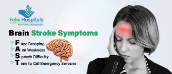
Recognizing the symptoms of a brain stroke is critical for getting prompt medical attention. The acronym F.A.S.T. is often used to remember the most common Brain stroke symptoms.

Subscribe to our
© Copyright 2025. All Rights Reserved by Felix Healthcare Private Limited
Design and development by :

 Emergency : +(91)9667064100