 WhatsApp

Subscribe to our
Some medical conditions develop slowly. They give warnings over months and years of rising cholesterol, creeping blood pressure, gradual breathlessness on exertion. You have time to catch them, time to act, time to course-correct.
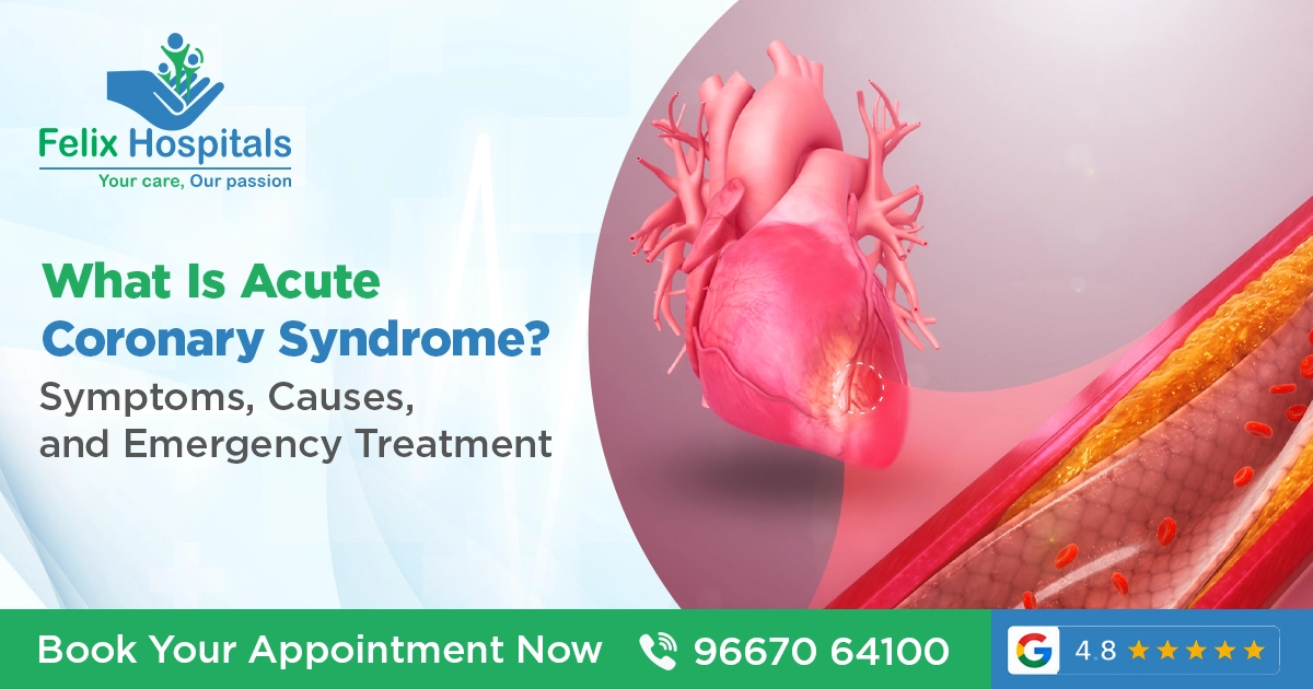
Acute Coronary Syndrome is not one of those conditions.
When it strikes, it strikes fast. The chest tightens, the arm goes numb, the cold sweat breaks out. What is happening inside the heart is a plaque rupturing, a clot forming, blood supply cutting off to heart muscle that cannot survive without it happening in real time. And what you do in the next thirty minutes to two hours determines whether that heart muscle lives or dies.
This is why Acute Coronary Syndrome is not simply a medical topic worth knowing about. It is information that can save a life of yours or someone standing next to you.
At Felix Hospital, one of the best cardiology hospitals in Noida, our cardiac emergency team manages Acute Coronary Syndrome with the urgency and precision this condition demands, around the clock. Our best cardiologists in Noida understand that in ACS, time is muscle and every minute of delay costs heart tissue that never grows back.
Here is everything you need to know.
Acute Coronary Syndrome (ACS) is an umbrella term covering a spectrum of life-threatening conditions that occur when blood supply to the heart muscle is suddenly, severely reduced or completely cut off.
The heart is a muscle and like all muscles, it needs a continuous supply of oxygen-rich blood to function. That blood arrives through the coronary arteries two main vessels (the left and right coronary arteries) that branch off the aorta immediately above the aortic valve and wrap around the surface of the heart, sending smaller branches into the heart muscle itself.
Under normal conditions, these arteries deliver blood smoothly and continuously. But in people with coronary artery disease where years of cholesterol deposition have built up plaques inside the arterial walls this delivery can be suddenly interrupted.
The interruption happens not gradually but acutely triggered most often by the rupture or erosion of a vulnerable plaque. When a plaque breaks open, the body's clotting system responds immediately platelets rush to the rupture site, fibrin is laid down, and a thrombus (blood clot) forms. If that clot is large enough, it narrows or completely blocks the artery.
When blood flow is severely reduced, heart muscle downstream is starved of oxygen. This is called myocardial ischaemia. When it is completely blocked for long enough heart muscle begins to die. This is called myocardial infarction, a heart attack.
Acute Coronary Syndrome is the collective name for this entire emergency spectrum. Where exactly on that spectrum a patient falls depends on the degree of arterial blockage, how long it has been present, and which artery is involved.
ACS is not a single diagnosis it is three distinct but related conditions, distinguished by ECG findings and blood test results. Understanding the difference matters because treatment is different for each.
This is the most severe form of ACS, the "classic" heart attack most people picture.
A coronary artery is completely blocked. Blood flow has stopped entirely to the section of heart muscle it supplies. The damage begins immediately and progresses with every passing minute. On the ECG, the ST segments of a specific portion of the cardiac waveform are elevated, indicating full-thickness involvement of the heart wall.
STEMI is the most time-critical of all cardiac emergencies. The treatment goal of reopening the blocked artery must be achieved within 90 minutes of hospital arrival for primary angioplasty (the preferred treatment), and within 30 minutes if clot-busting drugs are used instead.
Every minute of delay from symptom onset to artery reopening results in the death of approximately 1.9 million cardiac muscle cells. By 90 minutes, a significant proportion of the affected territory may be irreversibly damaged.
In NSTEMI, a coronary artery is severely but not completely blocked. Blood flow is critically reduced but not entirely absent. Heart muscle damage is occurring evidenced by elevation of cardiac biomarkers (troponin) in the blood but the ECG does not show the classic ST elevation pattern.
NSTEMI is slightly less immediately catastrophic than STEMI but only slightly. It still represents irreversible myocardial damage and carries significant short- and medium-term risk of progression to complete occlusion or sudden cardiac death. It requires urgent hospitalisation, antiplatelet and anticoagulant therapy, and typically coronary angiography within 24 to 72 hours.
In unstable angina, a vulnerable plaque has ruptured and a partially obstructing clot has formed but the artery is not completely blocked and the blood flow reduction has not yet caused measurable heart muscle damage. Troponin levels are normal. The ECG may show ST depression or T-wave changes, or may be entirely normal.
Unstable angina is essentially NSTEMI without the enzyme rise but it is an emergency nonetheless. It represents a heart on the edge. The clot that has not fully blocked the artery today can extend and block it completely tomorrow converting unstable angina into NSTEMI or STEMI without warning.
Patients with unstable angina must be admitted, monitored, and treated as an ACS not sent home with pain relief.
The Primary Cause: Atherosclerosis and Plaque Rupture
The overwhelming majority of ACS events, more than 90 percent, occur in the setting of atherosclerosis: the progressive accumulation of cholesterol-rich plaques within the walls of the coronary arteries.
This process begins early sometimes in the second and third decade of life and progresses silently for decades. The plaques narrow the coronary arteries progressively, but stable plaques often do not cause symptoms until the narrowing exceeds 70 percent of the vessel diameter. Even then, stable plaques may only cause symptoms of exertion which is called stable angina.
The critical event that triggers ACS is not gradual narrowing; it is plaque rupture or erosion. When the fibrous cap covering a vulnerable plaque breaks, the lipid-rich core of the plaque is exposed to circulating blood. This triggers the clotting cascade platelets to adhere, aggregate, and activate; fibrin threads form; and within minutes, a thrombus develops at the rupture site.
Paradoxically, the plaques most likely to rupture are not the largest or most obstructing ones they are soft, lipid-rich, thin-capped plaques that may have been causing only mild narrowing before the rupture. This is why coronary angiography done months before an ACS event can show only mild disease in the vessel that subsequently occludes the dangerous plaque was there but not causing significant obstruction.
While atherosclerotic plaque rupture is the dominant mechanism, ACS can also occur through:
Coronary artery spasm sudden intense contraction of the arterial wall that dramatically narrows or transiently closes the artery. Prinzmetal's (variant) angina is the classic example. Cocaine use can trigger severe coronary spasm even in young people with no atherosclerosis.
Spontaneous Coronary Artery Dissection (SCAD) a tear in the wall of the coronary artery that creates a false channel, compressing the true lumen. Seen disproportionately in young women, often without traditional cardiovascular risk factors. Associated with pregnancy, extreme physical stress, and connective tissue disorders.
Coronary embolism is a clot originating elsewhere (such as from a valve, the left atrium in atrial fibrillation, or a prosthetic valve) that travels to and lodges in a coronary artery.
Demand ischaemia in severely ill patients, extreme physiological stress (severe anaemia, sepsis, hypotension, or very fast heart rates) can outstrip the supply capacity of even mildly diseased coronary arteries, producing myocardial ischaemia and infarction without a new plaque event. This is called Type 2 MI.
ACS does not appear from nowhere. In almost every case, there are identifiable risk factors that have been silently building the conditions for an event over years.
These cannot be changed but knowing them helps in understanding and quantifying personal risk:
Age risk increases substantially after 45 in men and after 55 in women (or after menopause)
Male sex men have a significantly higher risk of ACS at younger ages; women's risk increases after menopause
Family history a first-degree relative (parent or sibling) who had a heart attack or coronary artery disease before age 55 (men) or 65 (women) significantly elevates personal risk
Ethnicity South Asians, including Indians, have a disproportionately high risk of coronary artery disease often occurring at younger ages and with more aggressive progression than in Western populations
These can be changed and changing them is the most powerful preventive medicine available:
Hypertension (high blood pressure) damages the arterial wall, accelerates atherosclerosis, and increases the mechanical stress on plaques
Dyslipidaemia elevated LDL ("bad") cholesterol drives plaque formation; low HDL ("good") cholesterol reduces the capacity to remove cholesterol from plaques
Diabetes mellitus dramatically accelerates atherosclerosis through multiple mechanisms; diabetic patients have a two- to four-fold higher risk of coronary events
Smoking one of the most potent cardiovascular toxins; directly damages endothelium, promotes clot formation, raises LDL, lowers HDL, and accelerates plaque progression
Obesity particularly central (abdominal) obesity drives insulin resistance, hypertension, and dyslipidaemia simultaneously
Physical inactivity sedentary lifestyle is an independent cardiovascular risk factor
Chronic psychological stress sustained stress elevates cortisol, promotes inflammation, drives hypertension, and is associated with plaque vulnerability
Poor diet high in trans fats, saturated fats, refined carbohydrates, and excess sodium; directly contributes to dyslipidaemia and hypertension
Excessive alcohol consumption elevates blood pressure and triglycerides
Cocaine and stimulant drug use causes coronary spasm, accelerated atherosclerosis, and arrhythmias
Chronic kidney disease an independent and under-recognised cardiovascular risk factor
Inflammatory conditions rheumatoid arthritis, psoriasis, systemic lupus erythematosus; chronic inflammation accelerates atherosclerosis
Obstructive sleep apnoea causes repeated nocturnal hypoxia, sympathetic activation, and blood pressure surges that damage the coronary vasculature
High-sensitivity CRP elevated inflammatory marker independently predicts coronary events beyond traditional risk factors
Lipoprotein(a) a genetically determined lipid particle; elevated Lp(a) is a significant and underdiagnosed cardiovascular risk factor
Recognising the symptoms of ACS and recognising them fast is the most important thing a layperson can do to save a life. Many people who die of heart attacks outside hospital do so not because treatment was unavailable but because they waited too long to seek it.
Chest pain or discomfort is the most common presenting symptom. Typically described as a pressure, heaviness, tightness, squeezing, or crushing sensation in the centre or left side of the chest. People frequently describe it as feeling like "a heavy weight sitting on the chest" or "a band tightening around the chest." It is not always sharp or stabbing; those descriptors are more typical of musculoskeletal or pleuritic pain.
Radiation of pain may radiate to the left arm (the most classic pattern), both arms, the jaw, the neck, the throat, or the upper back. Jaw pain that is otherwise unexplained in the context of exertion or rest should always raise cardiac suspicion.
Breathlessness difficulty breathing, often accompanying the chest discomfort or sometimes occurring without it; caused by the acutely impaired pump function of the ischaemic heart.
Diaphoresis cold, clammy sweating that is sudden and unexplained; a classic autonomic response to myocardial ischaemia that is highly suggestive of ACS.
Nausea and vomiting are particularly common in inferior wall myocardial infarctions (those affecting the bottom of the heart, supplied by the right coronary artery), due to vagal stimulation.
Dizziness and light-headedness from reduced cardiac output and blood pressure.
Palpitations awareness of irregular or fast heartbeat, ACS frequently triggers arrhythmias including ventricular fibrillation, which can cause sudden cardiac arrest.
Sense of impending doom a profound, overwhelming feeling that something catastrophic is happening; reported by many patients and considered a significant clinical indicator of serious cardiovascular events.
Not all ACS events present with dramatic chest pain. Atypical presentations are more common than most people realise and they are a major reason why ACS is missed or delayed in certain groups.
Women are significantly more likely to present without classic chest pain instead experiencing predominant fatigue, jaw pain, upper back pain, nausea, or breathlessness as the main symptom
Diabetic patients particularly those with longstanding diabetes frequently experience silent myocardial infarctions due to diabetic neuropathy affecting the cardiac sensory nerves; they may have no pain at all, presenting only with breathlessness or sudden fatigue
Elderly patients may present with confusion, syncope (fainting), or extreme weakness
Epigastric pain (upper abdominal pain, like severe indigestion) particularly in inferior wall MI is a classic ACS mimic that delays diagnosis
The message is this: chest discomfort that is new, unexplained, and accompanied by breathlessness, sweating, or radiation to the arm or jaw is ACS until proven otherwise. Do not self-diagnose as indigestion. Do not wait to see if it passes. Call for emergency help immediately.
When a patient arrives at Felix Hospital's emergency department with suspected ACS, the clock starts immediately. Diagnosis and treatment happen in parallel not sequentially.
The ECG is the single most important and time-critical investigation. It must be performed and interpreted within 10 minutes of the patient's arrival at the emergency department; this is an international standard.
ST elevation indicating STEMI; triggers the immediate activation of the cardiac catheterisation laboratory
ST depression or T-wave inversions suggesting NSTEMI or unstable angina
New left bundle branch block treated as a STEMI equivalent
Normal ECG does not rule out ACS; up to 5 percent of confirmed MIs have a normal initial ECG
High-sensitivity troponin (hs-cTnI or hs-cTnT) is the blood test that detects heart muscle damage. Troponin is a protein released into the bloodstream when cardiac muscle cells are injured or dying.
Modern high-sensitivity troponin assays can detect minute elevations within 1 to 3 hours of symptom onset. Serial troponin measurements at presentation and 1 to 3 hours later form the basis of the rapid ACS diagnostic protocol used internationally and at Felix Hospital.
A rising troponin level in the context of symptoms myocardial infarction. The pattern of rise and fall is as diagnostically important as the absolute level.
Chest X-ray assesses cardiac size and pulmonary oedema (fluid in the lungs indicating heart failure).
Echocardiography ultrasound of the heart; identifies regional wall motion abnormalities (sections of heart muscle not contracting normally) that correspond to the ischaemic territory, and assesses overall pump function (ejection fraction).
Full blood count, renal function, glucose, lipid profile to assess baseline organ function, identify co-morbidities, and guide medication choices.
Coronary angiography the definitive investigation: a catheter is passed through the femoral or radial artery under X-ray guidance into the coronary arteries, and contrast dye is injected to visualise the coronary anatomy, identify the culprit lesion, and guide treatment. In STEMI, this happens immediately as part of primary PCI (angioplasty). In NSTEMI, it typically occurs within 24 to 72 hours of admission.
Call emergency services immediately. Do not drive yourself. Do not wait for the pain to improve. Every minute without treatment is heart muscle dying.
Aspirin if the patient is not allergic to aspirin and can swallow safely, a single dose of 325mg aspirin (chewed, not swallowed whole, for faster absorption) given immediately reduces clot propagation. This is one of the few things that can be done before hospital arrival that meaningfully affects outcomes.
Position the patient comfortably, usually sitting up or semi-reclined; lying flat may worsen breathlessness.
Loosen any restrictive clothing.
If the patient is unconscious and not breathing normally begin CPR immediately and continue until emergency services arrive.
Oxygen administered if oxygen saturation is below 90 percent.
Intravenous access established immediately for medication delivery.
Continuous cardiac monitoring ECG monitoring detects life-threatening arrhythmias the moment they arise.
Antiplatelet therapy dual antiplatelet therapy is the cornerstone of ACS treatment:
Aspirin (if not already given)
P2Y12 inhibitor ticagrelor, prasugrel, or clopidogrel; these drugs block platelet activation and aggregation at the rupture site, preventing clot growth
Anticoagulation heparin (unfractionated or low molecular weight) is administered to prevent further clot formation.
Nitrates glyceryl trinitrate (GTN) sublingually or intravenously; dilates coronary arteries and reduces preload, relieving chest pain and reducing myocardial oxygen demand.
Beta-blockers slow the heart rate and reduce myocardial oxygen demand; started early when haemodynamically appropriate.
Morphine for pain relief and reduction of sympathetic activation in patients with ongoing severe pain; used judiciously as there is some evidence of interaction with oral antiplatelet agents.
Statins high-intensity statin therapy is started immediately; reduces inflammation at the plaque site and has early benefits beyond cholesterol lowering.
For patients with STEMI, primary PCI (coronary angioplasty) is the most effective treatment when performed within 90 minutes of hospital arrival, the international "door-to-balloon" time standard.
The procedure:
A thin, flexible catheter is introduced through the radial artery at the wrist (or femoral artery at the groin) under local anaesthesia. The catheter is guided through the arterial system under X-ray imaging to the blocked coronary artery. A guidewire is passed through the blockage. A balloon catheter is inflated at the site of the blockage compressing the clot and opening the artery. In most cases, a coronary stent, a small metal mesh tube, is then deployed at the site to keep the artery open and prevent recoil.
The results are immediate: the blocked artery is reopened, blood flow is restored, and heart muscle that was dying is salvaged.
At Felix Hospital, our cardiac catheterisation laboratory is equipped for 24-hour primary PCI delivery because heart attacks do not respect office hours.
When a patient presents to a hospital without an immediately available catheterisation laboratory, thrombolytic therapy (clot-busting drugs streptokinase, alteplase, tenecteplase) can restore coronary blood flow pharmacologically.
Thrombolysis must be administered within 12 hours of symptom onset and ideally within the first 3 hours for maximum benefit. It carries a small but definite risk of serious bleeding including intracranial haemorrhage and is therefore contraindicated in patients with recent surgery, stroke history, or uncontrolled hypertension.
Even after successful thrombolysis, patients should be transferred to a centre with angiography capability for evaluation within 3 to 24 hours, a strategy known as pharmacoinvasive approach.
In a minority of ACS patients those with complex multi-vessel coronary disease, left main stem disease, or anatomy unsuitable for PCI coronary artery bypass surgery is the preferred revascularisation strategy. This involves using arterial or venous grafts to bypass the blocked segments of coronary artery, restoring blood supply through new conduits.
In the acute setting, CABG is generally reserved for patients who cannot be managed by PCI or who have mechanical complications of MI (such as ventricular septal rupture or papillary muscle rupture).
Surviving the acute event is step one. The medications prescribed after an ACS event are not a temporary measure; they are lifelong protection against the next event.
Dual antiplatelet therapy (DAPT) aspirin combined with a P2Y12 inhibitor (ticagrelor or clopidogrel) for a minimum of 12 months after ACS, and indefinitely in some high-risk patients. This is the most important drug combination for preventing stent thrombosis and recurrent coronary events.
High-intensity statin atorvastatin 40 to 80mg or rosuvastatin 20 to 40mg daily; the target LDL is below 1.4 mmol/L (55 mg/dL) aggressive reduction that significantly reduces recurrence risk.
Beta-blocker reduces heart rate and blood pressure, decreases myocardial oxygen demand, and has specific survival benefits post-MI, particularly in patients with reduced ejection fraction.
ACE inhibitor or ARB reduces remodelling of the damaged heart muscle, lowers blood pressure, and is particularly important in patients with reduced ejection fraction or diabetes.
Aldosterone antagonist eplerenone or spironolactone; added in patients with significantly reduced ejection fraction after MI to reduce heart failure progression.
Proton pump inhibitor omeprazole or pantoprazole; used alongside DAPT to protect the stomach lining from the increased bleeding risk of dual antiplatelet therapy.
The first 24 to 48 hours after ACS are the period of highest risk for arrhythmias, haemodynamic deterioration, and mechanical complications.
Ventricular fibrillation (VF) and sudden cardiac death are the most feared immediate complications. VF chaotic electrical activity in the ventricles causes cardiac arrest within seconds. This is the primary reason why cardiac monitoring is continuous in the first 24 to 48 hours and why defibrillators are immediately available.
Acute heart failure and cardiogenic shock when a large area of heart muscle is damaged, the pump function of the heart fails catastrophically. Cardiogenic shock where blood pressure falls and organs begin to fail from inadequate perfusion carries a mortality of 40 to 50 percent even with aggressive treatment.
Mechanical complications rare but catastrophic events that can occur in the days following MI:
Ventricular septal defect rupture of the wall between the heart's lower chambers
Papillary muscle rupture causing acute severe mitral regurgitation
Free wall rupture rupture of the outer wall of the ventricle; almost universally fatal without immediate surgery
Left ventricular thrombus a blood clot forming inside the damaged ventricle; can embolise to cause stroke.
Pericarditis inflammation of the sac surrounding the heart; occurs in the days following MI (Dressler's syndrome is a later, autoimmune variant).
Recurrent ischaemia the culprit plaque site and other vulnerable plaques remain at risk; this is why long-term antiplatelet and statin therapy is non-negotiable.
Depression and anxiety experienced by a significant proportion of patients after ACS; independently associated with worse cardiovascular outcomes if not recognised and treated. Cardiac rehabilitation combining supervised exercise, education, and psychological support significantly reduces recurrence risk.
ACS prevention is not complicated. It is disciplined.
The most effective preventive measures all of which are within reach of most people target the modifiable risk factors that drive atherosclerosis and plaque vulnerability.
Control blood pressure. Target below 130/80 mmHg. Take prescribed medications without gaps. Monitor at home. Know your numbers.
Manage cholesterol. Get a fasting lipid profile done. Know your LDL level. If it is elevated, dietary changes and statins reduce cardiovascular events significantly. If you have already had an ACS event, your LDL target is below 55 mg/dL, considerably more aggressive than for primary prevention.
Stop smoking. No cardiovascular risk reduction strategy is more cost-effective or more immediately impactful than smoking cessation. Within 12 months of quitting, the excess cardiovascular risk from smoking is halved.
Control blood sugar. Diabetes and pre-diabetes dramatically accelerate coronary artery disease. HbA1c should be below 7 percent in most diabetic patients. Diet, exercise, and medication all contribute.
Move every day. 150 minutes of moderate aerobic exercise per week walking, swimming, cycling reduces cardiovascular event risk by 35 percent. It does not require a gym.
Eat a heart-protective diet. Mediterranean-style eating rich in vegetables, fruits, whole grains, olive oil, nuts, fish, and legumes has the strongest evidence base for cardiovascular protection. Reduce processed foods, refined carbohydrates, trans fats, and excessive sodium.
Maintain a healthy weight. BMI between 18.5 and 24.9, with waist circumference below 90cm in men and 80cm in women (for South Asians where the thresholds are lower than Western populations due to metabolic differences).
Manage stress. Chronic psychological stress is an independent cardiovascular risk factor. Yoga, meditation, regular exercise, adequate sleep, and social connection all contribute to stress resilience.
Attend regular cardiac check-ups. A cardiovascular risk assessment including blood pressure, fasting glucose, fasting lipids, BMI, and family history review should be part of every adult's annual health review, particularly after the age of 35 in South Asians.
This cannot be overstated. If you or someone near you experiences:
Chest pain, pressure, tightness, or heaviness especially if it lasts more than 5 minutes
Chest discomfort with sweating, nausea, or breathlessness
Pain radiating to the left arm, jaw, neck, or upper back
Sudden severe breathlessness without obvious cause
Loss of consciousness or collapse
Call emergency services immediately. Do not drive yourself to the hospital. Do not wait to see if symptoms improve. Do not self-treat with antacids hoping it is indigestion.
The window for maximum benefit from primary PCI in STEMI is 90 minutes from hospital arrival. The window for thrombolysis is 12 hours from symptom onset. Every minute of delay is measurable myocardial damage.
Felix Hospital, Sector 137, Noida is equipped to manage acute coronary syndrome as the true emergency is 24 hours a day, seven days a week, every day of the year.
Our cardiac emergency infrastructure includes:
24-hour cardiac emergency department with ECG performed and interpreted within 10 minutes of arrival
High-sensitivity troponin testing with rapid turnaround for ACS risk stratification
Cardiac catheterisation laboratory available for primary PCI in STEMI around the clock
Cardiac Intensive Care Unit (CICU) for continuous haemodynamic monitoring and arrhythmia management
Interventional cardiology team experienced in primary PCI, complex coronary intervention, and mechanical circulatory support
Cardiovascular surgery for patients requiring CABG or management of mechanical complications
Cardiac rehabilitation programme structured exercise, education, and psychological support post-ACS
Preventive cardiology outpatient clinic for cardiovascular risk assessment, lipid management, hypertension control, and secondary prevention
The best cardiologists in Noida are at Felix Hospital not as a claim, but as a commitment visible in outcomes, infrastructure, and the protocols our team follows every single day.
A heart attack technically myocardial infarction is one type of Acute Coronary Syndrome. ACS is the broader umbrella term that includes STEMI (complete arterial blockage with heart muscle death), NSTEMI (partial blockage with heart muscle damage), and unstable angina (partial blockage without detectable muscle damage). All three are emergencies. A heart attack refers specifically to STEMI and NSTEMI situations where heart muscle is dying or has died.
Within the first 20 minutes of complete coronary occlusion, heart muscle begins to die. The process accelerates over the following hours. By 6 hours without reperfusion, the majority of the myocardium supplied by the blocked artery may be irreversibly infarcted. This is why the phrase "time is muscle" is not a cliché, it is a physiological fact.
Yes. While ACS predominantly affects people over 45 to 50, it occurs in younger adults particularly those who smoke, have diabetes, have a strong family history of premature coronary disease, or use cocaine. South Asians tend to develop coronary artery disease at younger ages than Western populations. ACS in people under 40 is uncommon but not rare.
Stable angina is chest discomfort brought on predictably by exertion and relieved by rest or nitrates within minutes caused by a stable plaque narrowing a coronary artery enough to restrict blood flow during increased demand. ACS is caused by an acute plaque rupture and clot formation occurring at rest or with minimal exertion, not relieved by rest, and representing a fundamentally different and far more dangerous physiological event.
Yes and this is critically important. Women, diabetic patients, and elderly individuals frequently experience ACS without classic chest pain. The presenting symptoms may be fatigue, breathlessness, jaw pain, upper back pain, nausea, or sudden sweating. Any combination of these symptoms particularly in a person with cardiovascular risk factors warrants urgent medical evaluation.
Without treatment, STEMI carries a 30-day mortality of 30 to 50 percent. Even those who survive untreated or undertreated ACS are left with significant heart muscle damage leading to chronic heart failure, arrhythmias, and dramatically reduced quality of life and life expectancy. There is no such thing as "waiting it out" with a heart attack.
Most patients are discharged from hospital within 3 to 5 days after uncomplicated STEMI treated with primary PCI. Full recovery returning to normal activities including work and exercise typically takes 4 to 6 weeks for uncomplicated cases. Patients with larger infarctions, reduced ejection fraction, or complications have longer recovery periods. Cardiac rehabilitation, a supervised exercise and education programme significantly accelerates and improves recovery.
Yes for the core medications. Aspirin is typically lifelong. The P2Y12 inhibitor is given for a minimum of 12 months, sometimes longer. Statins and ACE inhibitors are indefinite in most patients. These medications are not treating symptoms, they are reducing the risk of the next event, which is a permanent biological risk that does not disappear after the acute episode is treated.
Call +91 9667064100 the line is answered 24 hours a day. Felix Hospital is located at Sector 137, Noida. For suspected cardiac emergencies, a call before coming our team can begin pre-arrival preparation that reduces door-to-balloon time.
A stent procedure can be life-saving for those with blocked arteries. This guide walks you through the stent experience—from the angioplasty process to post-procedure recovery and lifestyle changes.
Find the best cardiologist for your heart care with these essential tips and expert advice.
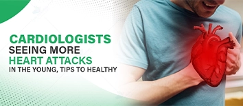
However, stress, sedentary lifestyle, chronic diseases such as diabetes and hypertension, and genetic factors place young individuals at risk of heart attack.

Top cardiologist surgeon in Noida for safe and expert pacemaker implantation to regulate heart rhythm.

Subscribe to our
© Copyright 2026. All Rights Reserved by Felix Healthcare Private Limited
Design and development by :


 +(91)9667064100
 Emergency : +(91)9667064100