 WhatsApp

Subscribe to our
Most people have heard of a heart attack. Most people know what a stroke is. But endocarditis, an infection of the heart's inner lining is something that flies completely under the public radar, often until it has already done serious damage.
Endocarditis can destroy a heart valve within days. It can send infected clots to the brain, the kidneys, and the lungs. It can mimic a dozen other illnesses for weeks before anyone suspects the heart is involved. And by the time it is correctly identified in patients who are delayed, the window for straightforward treatment has often already closed.
At Felix Hospital, recognized as one of the best cardiology hospitals in Noida, our team of senior cardiologists and cardiovascular surgeons manages infective endocarditis with the urgency and precision this condition demands. Our best cardiologists in Noida understand that endocarditis is not a condition that waits and neither should the people who have it or are at risk of it.
This guide covers everything about what endocarditis is, what it looks like, who is most vulnerable, how it is diagnosed and treated, and what happens when it is missed.
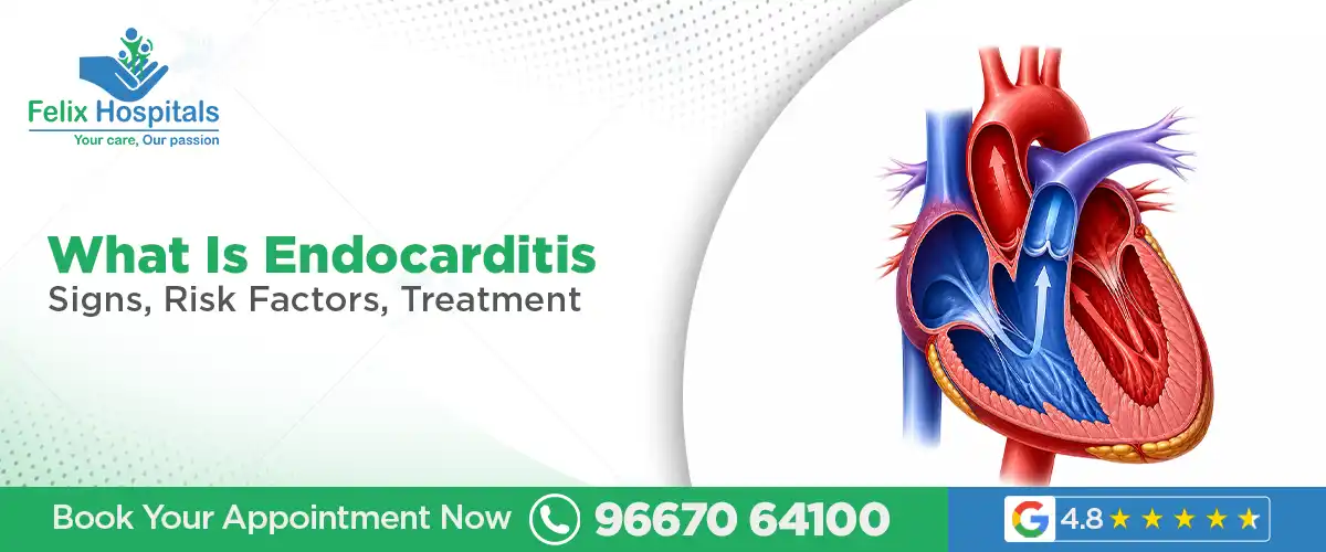
The heart is lined on the inside by a thin, smooth layer of tissue called the endocardium. It covers the inner walls of the heart chambers and, critically, the surfaces of the four heart valves, the mitral, aortic, tricuspid, and pulmonary valves which open and close with every heartbeat to keep blood moving in the right direction.
Endocarditis is inflammation of the inner lining almost always caused by infection.
When bacteria, fungi, or other microorganisms enter the bloodstream and reach the heart, they can adhere to the endocardial surface particularly to the valves, where the mechanical stress of constant opening and closing creates small irregularities that act as attachment sites. Once attached, they multiply and trigger an immune response. The body's attempt to fight the infection produces deposits of platelets, fibrin, and bacteria clumped together called vegetations on the valve surface.
These vegetations are the hallmark of infective endocarditis. They are not benign growths. They interfere with valve function, they can break off and travel through the bloodstream as infected emboli causing strokes, kidney damage, or abscesses anywhere in the body and they progressively damage and destroy the valve they sit on.
Left untreated, infective endocarditis is almost universally fatal. Even with treatment, it remains one of the most serious infections in cardiovascular medicine carrying an in-hospital mortality of 15 to 30 percent in many published series.
Endocarditis is not a single, uniform disease. It presents differently depending on the causative organism, the speed of onset, and the underlying condition of the heart valves.
This is the most common and clinically significant form. It is caused by a microbial infection, most often bacterial. Infective endocarditis is further categorized by its course:
Acute Infective Endocarditis Caused by highly virulent organisms most commonly Staphylococcus aureus (including MRSA). It progresses rapidly over days destroying valves, causing high fever, sepsis, and haemodynamic deterioration within a short time. Acute IE is a medical emergency from day one.
Subacute Infective Endocarditis (SBE Subacute Bacterial Endocarditis) Caused by less virulent organisms classically Streptococcus viridans, which normally lives harmlessly in the mouth. It progresses slowly over weeks to months with insidious, low-grade symptoms that mimic many other conditions. This is the form that is most frequently diagnosed late, because for weeks it may look like a viral illness, fatigue syndrome, or early rheumatic disease.
Less common but worth understanding:
Libman-Sacks Endocarditis associated with systemic lupus erythematosus (SLE). Non-infective vegetations form on both surfaces of the mitral valve. Not caused by bacteria but can mimic infective endocarditis.
Marantic (Thrombotic) Endocarditis seen in patients with advanced cancer or other chronic debilitating illnesses. Sterile vegetations form on valves, often as a paraneoplastic phenomenon, and can embolise.
Rheumatic Endocarditis a consequence of acute rheumatic fever following streptococcal throat infection. Inflammation of the valve leaflets particularly the mitral valve can lead to long-term scarring and rheumatic heart disease. Historically a leading cause of heart valve disease in India.
A distinct and particularly serious category endocarditis occurring on an artificial (prosthetic) heart valve. It is divided into:
Early PVE occurring within 60 days of valve surgery; usually caused by organisms introduced at the time of surgery
Late PVE occurring after 60 days; pathogenesis similar to native valve endocarditis
Prosthetic valve endocarditis is harder to treat, more likely to require surgery, and carries a higher mortality than native valve disease.
Left-sided endocarditis (affecting the mitral and aortic valves) is far more common and generally more dangerous because these valves handle the systemic circulation.
Right-sided endocarditis (affecting the tricuspid valve) is strongly associated with intravenous drug use and typically presents with pulmonary complications septic pulmonary emboli, lung abscesses rather than systemic embolisation.
The root cause of infective endocarditis is always the same: microorganisms entering the bloodstream (a process called bacteraemia) and successfully colonising the heart's inner surfaces.
Bacterial causes the overwhelming majority:
Staphylococcus aureus the most common cause of acute endocarditis globally; associated with healthcare-related infections, IV drug use, skin infections, and indwelling catheters
Streptococcus viridans group historically the most common cause of subacute endocarditis; bacteria that normally inhabit the mouth and enter the bloodstream during dental procedures or poor oral hygiene
Enterococcus faecalis associated with gastrointestinal and urinary tract procedures; often affecting older adults
Streptococcus bovis (gallolyticus) associated with colorectal polyps and colon cancer; a diagnosis of endocarditis with this organism should prompt colonoscopy
HACEK group organisms (Haemophilus, Aggregatibacter, Cardiobacterium, Eikenella, Kingella) slow-growing mouth bacteria causing a small but recognised proportion of cases
Coagulase-negative staphylococci particularly relevant in prosthetic valve endocarditis
Fungal endocarditis: Far less common but significantly more difficult to treat. Caused by Candida and Aspergillus species most often in immunocompromised patients, those on prolonged IV nutrition, or those with prosthetic valves. Carries an extremely high mortality.
Bacteraemia the presence of bacteria in the bloodstream occurs more frequently than most people realise:
Dental procedures particularly those involving manipulation of gum tissue
Routine daily activities in people with poor oral hygiene even vigorous toothbrushing or chewing food can cause transient bacteraemia
Urological procedures catheterisation, cystoscopy
Gastrointestinal endoscopy with biopsy
Skin infections boils, cellulitis, infected wounds
Intravenous drug injection using non-sterile needles introduces skin bacteria directly into the bloodstream
Indwelling venous catheters, pacemaker leads, dialysis access
Respiratory tract procedures
In most people, transient bacteraemia is cleared by the immune system without any consequence. In people with vulnerable hearts damaged valves, prosthetic valves, congenital abnormalities the bacteria find a foothold and the infection takes hold.
Not everyone who develops bacteraemia gets endocarditis. The risk is substantially concentrated in specific groups whose hearts are more susceptible to colonisation.
High-risk cardiac conditions:
Prosthetic heart valves mechanical or bioprosthetic; the surface of an artificial valve is a more hospitable attachment site for bacteria
Previous episode of infective endocarditis prior infection leaves scarring that increases the risk of recurrence significantly
Congenital heart disease particularly unrepaired cyanotic defects, repaired defects with residual abnormalities near prosthetic material, and specific repaired defects in the first 6 months after surgery
Rheumatic heart disease scarred, damaged valves from prior rheumatic fever are more vulnerable; this is particularly relevant in India, where rheumatic heart disease remains common
Degenerative valve disease calcific aortic stenosis and mitral valve prolapse with regurgitation
Bicuspid aortic valve congenital two-leaflet valve with abnormal blood flow dynamics
Lifestyle and exposure-related risk factors:
Intravenous drug use the single most significant lifestyle risk factor; introduces skin flora directly into the bloodstream repeatedly
Poor dental hygiene chronic periodontal disease creates sustained, low-level bacteraemia
Indwelling intravascular devices central venous catheters, haemodialysis catheters, pacemaker and defibrillator leads
Recent dental or surgical procedures in high-risk individuals
Immune and systemic risk factors:
Diabetes mellitus impairs immune response and increases susceptibility to infection generally.
Chronic kidney disease on dialysis access creates repeated opportunities for bacteraemia.
HIV infection with low CD4 counts reduced immune competence.
Chronic liver disease and cirrhosis.
Immunosuppressive therapy post-transplant patients, those on steroids or biological agents.
Advanced age immune function declines with age; degenerative valve disease is more common.
Endocarditis is one of medicine's great mimics. It can look like the flu, like rheumatoid arthritis, like a urinary tract infection, like a stroke. This mimicry is the reason diagnosis is so frequently delayed and why index of suspicion matters enormously in any patient with unexplained fever and a cardiac risk factor.
Fever is the most consistent symptom; present in the vast majority of patients. In acute endocarditis, it is high and spiking. In subacute endocarditis, it may be low-grade 37.5 to 38.5°C for weeks, with night sweats and chills. The persistence of fever that does not resolve with routine treatments should always raise alarm.
Constitutional symptoms include fatigue, malaise, generalised weakness, poor appetite, and unintentional weight loss; particularly prominent in subacute presentations.
Heart murmur a new or changing heart murmur, caused by the vegetation disrupting normal valve function, is a critical clinical finding. A new regurgitant murmur in a febrile patient with a cardiac risk factor is endocarditis until proven otherwise.
Joint and muscle pain arthralgia and myalgia are common, which is why endocarditis is so frequently confused with viral illness or rheumatological conditions in its early stages.
These are findings caused by immune complex deposition and septic or aseptic emboli affecting small vessels throughout the body. They are seen in subacute endocarditis more than acute.
Petechiae are small, pinpoint red spots that do not blanch on pressure; found on the skin, the conjunctivae (whites of the eyes), and the mucous membranes of the mouth. One of the more common peripheral signs.
Splinter haemorrhages thin, linear reddish-brown streaks under the fingernails or toenails, resembling splinters trapped beneath the nail. Caused by tiny emboli in the capillaries of the nail bed. Seen in up to 15 percent of patients.
Osler's nodes have small, painful, tender lumps on the pads of the fingers and toes; caused by immune complex deposition. Their tenderness distinguishes them from Janeway lesions. Classic but not commonly seen in approximately 10 to 25 percent of cases.
Janeway lesions are flat, irregular, painless red or haemorrhagic spots on the palms of the hands and soles of the feet; caused by septic emboli. More common in acute Staphylococcus aureus endocarditis.
Roth spots oval, white-centred haemorrhages visible on the retina on eye examination; caused by immune complex vasculitis. Seen in approximately 5 percent of patients but highly specific when present.
Clubbing of fingers seen in longstanding endocarditis; indicates chronic disease.
Splenomegaly an enlarged spleen, palpable on abdominal examination; common in subacute endocarditis because the spleen is chronically filtering septic material from the bloodstream.
In many patients particularly those with acute or advanced endocarditis the presenting complaint is not fever or malaise, but a complication:
Stroke or TIA presenting as sudden weakness, speech disturbance, or facial drooping; caused by cerebral embolisation from cardiac vegetations
Sudden visual loss from retinal artery embolism
Flank pain and haematuria from renal infarction
Sudden severe chest pain and breathlessness from acute heart failure due to valve destruction, or from pulmonary embolism in right-sided disease
Back pain from vertebral osteomyelitis seeded by septic emboli
Abdominal pain from splenic infarction or mesenteric embolism
Diagnosis of endocarditis rests on two pillars: blood cultures and echocardiography formalised in the internationally used Modified Duke Criteria, which combines clinical, microbiological, and imaging findings to classify cases as definite, possible, or rejected endocarditis.
Blood cultures are the single most important investigation. At least three sets of blood cultures from different venepuncture sites, at different times, should be drawn before any antibiotics are started. This matters enormously: blood cultures taken after antibiotics have been started are frequently negative, leaving the clinician treating blindly without knowing which organism is responsible or which antibiotics it is sensitive to.
Echocardiography:
Transthoracic Echocardiography (TTE) an ultrasound of the heart done through the chest wall. Useful for initial assessment and detecting large vegetations, valve dysfunction, and complications like pericardial effusion. Sensitivity is lower around 60 to 70 percent for small vegetations.
Transoesophageal Echocardiography (TOE/TEE) an ultrasound probe passed into the oesophagus gives far superior views of the heart valves particularly the mitral valve and prosthetic valves. Sensitivity exceeds 90 percent. TOE is recommended in virtually all clinically suspected cases, particularly when TTE is negative but suspicion remains high, when prosthetic valves are involved, or when complications are suspected.
Full blood count typically shows elevated white cell count (leucocytosis), anaemia of chronic disease, and elevated inflammatory markers (CRP, ESR).
Blood tests elevated inflammatory markers (CRP, ESR, procalcitonin), anaemia, elevated creatinine (if renal involvement), haematuria on urinalysis.
Chest X-ray may show pulmonary infiltrates in right-sided endocarditis, cardiomegaly, or pulmonary oedema from valve dysfunction.
CT scan CT of the chest, abdomen, and brain is increasingly performed to look for embolic complications, silent cerebral infarcts, splenic infarcts, renal infarcts, or mycotic aneurysms. CT angiography of the aorta may be needed to assess for periannular extension of infection.
PET-CT and Cardiac CT are newer imaging modalities that have significantly improved diagnostic sensitivity, particularly for prosthetic valve endocarditis and cardiac device infections, where echocardiography alone is less reliable.
Dental review is often requested to identify the source of bacteraemia, particularly in streptococcal endocarditis.
Endocarditis requires prolonged, high-dose intravenous antibiotic therapy this is not a condition treated with a short course of oral antibiotics. It is managed in hospital, with close monitoring of clinical response, cardiac function, and complications.
The duration of treatment depends on the organism, the valve involved, and whether native or prosthetic valve disease is present:
Streptococcal native valve endocarditis 4 weeks of IV penicillin or ceftriaxone; may be shortened to 2 weeks with the addition of gentamicin in uncomplicated cases with sensitive organisms
Staphylococcus aureus native valve endocarditis 4 to 6 weeks of IV flucloxacillin (or vancomycin if MRSA)
Prosthetic valve endocarditis minimum 6 weeks of IV antibiotics; regimens are more complex and often involve combination therapy
Enterococcal endocarditis 4 to 6 weeks of combination therapy (typically ampicillin plus gentamicin, or ampicillin plus ceftriaxone)
Fungal endocarditis IV antifungal therapy (amphotericin B or echinocandins); almost always requires surgical valve replacement as antifungals alone are rarely curative
Culture-negative endocarditis when blood cultures are negative (most commonly because antibiotics were given before cultures were taken, or due to fastidious organisms) empirical broad-spectrum therapy is used while further diagnostic workup is pursued.
Surgery is required in a significant proportion of endocarditis patients approximately 40 to 50 percent during the acute hospitalization phase. The decision to operate, and its timing, is one of the most critical and nuanced judgements in cardiovascular medicine.
Indications for surgery in infective endocarditis:
Heart failure caused by severe valve regurgitation or obstruction from large vegetations; the most common surgical indication and generally requires urgent or emergency operation
Uncontrolled infection persistent fever and positive blood cultures despite appropriate antibiotic therapy; suggests a paravalvular abscess, resistant organism, or fungal infection
Prevention of embolism very large vegetations (greater than 10 mm), particularly on the mitral valve, in patients who have already had one embolic event, carry high enough risk of recurrent embolism that surgery is warranted
Paravalvular extension abscess formation around the valve annulus; indicates invasive disease and often necessitates extensive reconstruction
Prosthetic valve endocarditis particularly early PVE or cases with paravalvular leak or dehiscence
Surgical procedures may include:
Valve repair when the valve structure allows, repair is preferred over replacement; avoids the need for lifelong anticoagulation with mechanical prostheses
Valve replacement mechanical or bioprosthetic valve; choice depends on patient age, anticoagulation tolerance, and other factors
Extensive debridement of infected and necrotic tissue, particularly in paravalvular abscess
Pacemaker or defibrillator lead removal when cardiac device infection is involved
The timing of surgery whether to operate in the acute phase of active infection or to delay is individualised. Early surgery (within the first week) is associated with better outcomes when heart failure or uncontrolled infection is present. A delay risks further valve destruction and further embolic events.
Beyond the primary antibiotic regimen, several other medications play important roles in management:
Anticoagulants the role of anticoagulation in endocarditis is complex and controversial. Patients with mechanical prosthetic valves who are already on anticoagulation present a particular challenge stopping anticoagulation risks valve thrombosis, but continuing it during active endocarditis increases the risk of haemorrhagic transformation of cerebral emboli. Decisions are made individually, by the treating team.
Diuretics in patients with heart failure due to valve regurgitation, diuretics (furosemide) and vasodilators help manage fluid overload and reduce cardiac preload while decisions about surgery are made.
Vasopressors in patients with septic shock which can occur in acute, aggressive endocarditis vasopressors (noradrenaline) are used to maintain blood pressure in the intensive care unit.
Antifungal agents echinocandins (anidulafungin, micafungin) and amphotericin B for fungal endocarditis.
Analgesics and antipyretics for symptomatic relief of fever, joint pain, and general malaise.
Rifampicin added to the antibiotic regimen in prosthetic valve endocarditis after the bacteraemia is controlled; improves biofilm penetration.
Endocarditis does not plateau. Without treatment, it progresses and its complications are severe, irreversible, and frequently fatal.
Embolic stroke is one of the most feared and common complications. Vegetations on left-sided valves the mitral and aortic are in the systemic circulation. When fragments break off, they travel to whatever artery is in their path most catastrophically, the cerebral arteries. Stroke occurs in 15 to 35 percent of endocarditis patients. It can be haemorrhagic (due to a mycotic aneurysm rupturing) or ischaemic (due to vessel occlusion). The resulting neurological damage may be permanent.
Acute heart failure develops when valve destruction reaches a tipping point the valve can no longer close properly, causing severe regurgitation. The heart, unable to compensate, fails acutely. In the context of active infection, this is a haemodynamic emergency that carries very high mortality without emergency surgery.
Paravalvular abscess infection extending beyond the valve into the surrounding tissue represents invasive, aggressive disease. It can cause complete heart block (when it involves the conducting system), fistula formation between cardiac chambers, and structural destruction that makes valve repair far more complex.
Mycotic aneurysms weaken spots in arterial walls caused by septic emboli seeding the vessel wall, forming aneurysms that can rupture. When this occurs in the brain a cerebral mycotic aneurysm rupture causes subarachnoid haemorrhage.
Renal failure from immune complex deposition in the kidney (glomerulonephritis), from septic renal emboli causing infarction, or from drug nephrotoxicity (aminoglycosides). Renal failure significantly worsens prognosis.
Septic emboli to other organs spleen (infarction, abscess), liver, mesentery (causing severe abdominal pain), and bone (vertebral osteomyelitis causing back pain that may be the presenting complaint in some patients).
Cardiac conduction abnormalities new heart block or bundle branch block developing during endocarditis treatment signals paravalvular extension and is an indication for urgent surgical reassessment.
Death in-hospital mortality ranges from 15 to 30 percent in published series, with higher rates in staphylococcal endocarditis, prosthetic valve involvement, elderly patients, and those with embolic complications.
Prevention of endocarditis is a conversation that belongs in every cardiology outpatient clinic because the people most at risk are identifiable, and the preventive measures are largely straightforward.
Current international guidelines recommend antibiotic prophylaxis before certain dental procedures specifically for patients in the highest-risk cardiac groups those with prosthetic heart valves, a previous episode of infective endocarditis, certain congenital heart defects, and cardiac valve disease in heart transplant recipients.
The regimen is typically a single oral dose of amoxicillin (or clindamycin if penicillin-allergic) taken 30 to 60 minutes before the procedure. It is important to understand that prophylaxis is not recommended before every dental visit; it is reserved for procedures involving manipulation of the gingival (gum) tissue or the periapical region of the teeth.
If you are in a high-risk cardiac group, your cardiologist at Felix Hospital will advise you on when prophylaxis is appropriate. This conversation should happen proactively not the morning before a dental appointment.
For people with high-risk hearts, oral hygiene is not just a cosmetic concern. Chronic periodontal disease creates sustained, low-level bacteraemia every time the person chews. Regular brushing, flossing, and six-monthly dental check-ups reduce the bacterial burden in the mouth and the frequency of oral bacteraemia significantly.
Encouraging patients with valvular heart disease to maintain excellent oral hygiene is one of the most evidence-based endocarditis prevention strategies available. It costs nothing and requires only habit.
Right-sided endocarditis caused by intravenous drug use is, in many settings, the fastest-growing subgroup of endocarditis cases. The repeated introduction of skin bacteria directly into the bloodstream with non-sterile needles is an almost direct route to tricuspid valve infection.
Cessation of IV drug use eliminates this risk. For individuals who are not yet able to stop, harm reduction programmes, clean needle exchange, supervised injection facilities, addiction treatment have a documented impact on endocarditis incidence and are a public health priority.
Any break in the skin wounds, ulcers, skin infections is a potential entry point for bacteria. Prompt treatment of skin infections, proper wound hygiene, and not allowing skin infections to be dismissed or self-treated inadequately reduces bacteraemia risk, particularly in high-risk patients.
Patients with indwelling venous catheters, pacemakers, or defibrillators should have these managed with strict aseptic technique at every interaction. Lines that are no longer necessary should be removed. Pacemaker lead infections are a specific and serious subtype of device-related endocarditis that requires complete lead extraction a complex procedure.
Patients with known valve disease including those with rheumatic heart disease, bicuspid aortic valve, or mitral valve prolapse with regurgitation should be under regular cardiology follow-up to monitor valve function over time. A valve that was mildly abnormal five years ago may be significantly more vulnerable today.
Regular echocardiography, guided by clinical assessment, allows the cardiologist to track progression and intervene surgically with a valve repair or replacement done electively before the valve becomes so diseased that it is one bacteraemia away from endocarditis.
At Felix Hospital, our cardiologists conduct structured valve disease follow-up programmes ensuring that patients with known cardiac abnormalities are not lost to follow-up in the years between significant clinical events.
Felix Hospital, Sector 137, Noida is recognised as one of the best cardiology hospitals in Noida with a full-service cardiac unit equipped to diagnose and manage infective endocarditis and its complications.
Our team of best cardiologists in Noida brings together the expertise of senior interventional cardiologists, cardiovascular surgeons, infectious disease specialists, and imaging experts because endocarditis is a condition that no single specialist can manage in isolation. It demands a team.
Our cardiology facilities include:
24-hour cardiac emergency services with round-the-clock echocardiography capability
Transoesophageal Echocardiography (TOE) the gold standard for endocarditis imaging
Cardiac CT and advanced imaging for embolic complication assessment and prosthetic valve evaluation
Cardiac Intensive Care Unit (CICU) for haemodynamically unstable patients
Cardiovascular Surgery for valve repair, valve replacement, and lead extraction
Microbiology and infectious disease for culture-guided antibiotic management and complex infection protocols
Outpatient cardiology for valve disease surveillance, antibiotic prophylaxis counselling, and endocarditis risk assessment
Whether you are experiencing symptoms that concern you, living with a high-risk cardiac condition and looking for a specialist to manage it proactively, or recovering from an endocarditis episode and needing ongoing cardiac care the team at Felix Hospital is here.
Call Felix Hospital: +91 9667064100
Endocarditis is an infection of the inner lining of the heart most critically, the heart valves. Bacteria or fungi that enter the bloodstream settle on the valves, multiply, and cause progressive damage. Without treatment, it is fatal. With prompt, appropriate treatment, most patients survive though recovery is prolonged and serious complications remain possible.
Bacteria enter the bloodstream a process called bacteraemia from many common sources: dental procedures, skin infections, urinary tract procedures, IV drug use, or indwelling catheters. In most people, the immune system clears them. In people with abnormal heart valves or prosthetic valves, the bacteria can attach to the damaged surface and start an infection.
Persistent unexplained fever particularly lasting more than a week without a clear cause in someone with a known heart valve abnormality, a prosthetic valve, or a history of endocarditis is the most important red flag. New or changing heart murmur, fatigue, night sweats, and any of the peripheral signs (splinter haemorrhages, Osler's nodes, petechiae) should prompt urgent cardiological evaluation.
Intravenous antibiotic treatment runs for 4 to 6 weeks, depending on the organism and whether native or prosthetic valves are affected. This is the minimum required to fully sterilise the infected valve. Shortening treatment increases the risk of relapse.
No approximately 50 to 60 percent of patients are managed with antibiotics alone. Surgery is required when heart failure develops due to valve destruction, when infection cannot be controlled with antibiotics, when the vegetations are very large and pose a high embolism risk, or when there is paravalvular abscess. These decisions are made by a multidisciplinary team on an individual basis.
Yes recurrence is a recognised risk, particularly in patients who continue IV drug use, those with prosthetic valves, and those with residual valve damage after a first episode. This is why long-term follow-up with a cardiologist, strict oral hygiene, and antibiotic prophylaxis before high-risk dental procedures are so important after an endocarditis episode.
Current guidelines recommend prophylaxis before certain dental procedures specifically for the highest-risk groups of patients with prosthetic heart valves, a prior episode of endocarditis, certain unrepaired congenital heart defects, and cardiac valve disease in transplanted hearts. If you are unsure whether you fall into this group, consult the cardiologists at Felix Hospital.
Call Felix Hospital at +91 9667064100 to book an outpatient cardiology consultation. Felix Hospital is located at Sector 137, Noida equipped with advanced cardiac imaging and a senior cardiology team experienced in managing valve disease and infective endocarditis.
Dr. Rahul Arora is an experienced Interventional Cardiologist with 21+ years of expertise in advanced cardiac care, specializing in personalized, evidence-based treatment for heart diseases and improved patient outcomes.
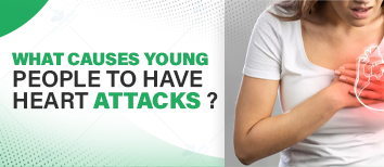
Heart disease is a significant health concern for women, often presenting unique challenges. Understanding the causes, symptoms, and treatment options is essential for early detection and effective management.
Learn how a CT Angiogram helps assess heart disease and your heart score effectively.

Subscribe to our
© Copyright 2026. All Rights Reserved by Felix Healthcare Private Limited
Design and development by :


 +(91)9667064100
 Emergency : +(91)9667064100